


| Journal of Current Surgery, ISSN 1927-1298 print, 1927-1301 online, Open Access |
| Article copyright, the authors; Journal compilation copyright, J Curr Surg and Elmer Press Inc |
| Journal website http://www.currentsurgery.org/ |
Review
Volume 5, Number 2-3, September 2015, pages 151-156
Parastomal Hernia Is a Problem Yet to Be Solved
Hagar Mizrahia, c, Nissim Gerona, Michael C. Parkerb
aDepartment of General Surgery, The Baruch Padeh Medical Center Poriya, Israel
bRoyal College of Surgeons, London, UK
cCorresponding Author: Hagar Mizrahi, Department of General Surgery, The Baruch Padeh Medical Center Poriya, Israel
Manuscript accepted for publication June 23, 2015
Short title: Repair of Parastomal Hernia
doi: http://dx.doi.org/10.14740/jcs268w
- Abstract
- Introduction
- Classification and Diagnosis of PH
- Risk Factor for the Development of PH
- Prevention of PH
- Clinical Manifestations of PH
- The Treatment of PH
- The Surgical Approach for the Repair of PH
- References
| Abstract | ▴Top |
Parastomal hernia is a common surgical problem which is associated with discomfort and irritation in addition to acute complications. This review presents the history, different classifications and terms which are used to describe parastomal herniation. It also details the different techniques for parastomal hernia diagnosis and presents evidence-based risk factors associated with its occurrence. It gives a detailed overview of modern treatment options and discusses different strategies for repair of parastomal hernia as well as advantages and disadvantages for each technique.
Keywords: Colostomy; Ileostomy; Parastomal; Hernia
| Introduction | ▴Top |
In surgical practice there is almost a daily necessity to fashion a diverting stoma with the earliest documented stoma created in 1710 by Alexis Littre [1]. Most stomas are formed as a temporary measure, but some are permanent. The most frequent complication of stoma creation is parastomal hernia (PH) which is defined as an incisional hernia related to a stoma site. Goligher is often cited for the statement that PH formation is an almost inevitable consequence of stoma formation [2].
| Classification and Diagnosis of PH | ▴Top |
The revised classification of Devlin and Kingsnorth for PH includes four subtypes: interstitial with a hernial sac within the muscle and aponeurotic layers of the abdomen, subcutaneous with a subcutaneous hernial sac, intrastomal in ileostomies with a hernial sac between the intestinal wall and the everted intestinal layer and peristomal with the bowel prolapsing through a circumferential hernial sac enclosing the aperture also known as prolapsed stoma [3]. Recently, other classifications were offered using clinical as well as other diagnostic methods in order to differentiate types of PH [4]. These diverse classifications represent not only terminological differences but signify the complexity of estimating the precise incidence of PH which varies between 5% and 52.0% of all stoma formations [2].
As a rule, PH diagnosis is made by physical examination. The patient needs to be examined in an erect and supine position with the abdomen exposed. Frequently a bulge is noticed which is more evident if the patient coughs or performs straight leg rising in the supine position. The examiner’s hand is laid near the stoma site to enable the impulse to be felt. When there is a doubt additional imaging tests can be performed [5, 6]. The use of imaging tools has increased and small PH are being discovered as an incidental finding in a CT performed for a different reason. Nevertheless, any attempt to rule out a diagnosis of PH should be delayed for at least 12 months after its creation, as some authors stress the importance of a prolonged post-operative period before estimating PH occurrence [2].
| Risk Factor for the Development of PH | ▴Top |
The risk factors related to PH occurrence are listed in Table 1. Similar to other incisional hernias, the risk for the development of PH is influenced by several factors which can be divided into two groups: patient-related and surgical-related factors. Among the patient-related factors age was proved as a risk factor in the Mylonakis et al study which demonstrated 22% of PH occurrence in patients older than 60 years compared with only 4.8% in younger patients (P = 0.02) [7]. This might be explained by a decrease of muscle thickness associated with ageing [8]. Patient’s habitus as formulated in obesity and abdominal perimeter was also proved to increase the rate of PH. De Raet and his co-workers offered a waist circumference of 100 cm as a threshold for the usage of prophylactic mesh during colostomy formation [9]. Pre-operative conditions such as emergency surgery, diabetes mellitus, ulcerative colitis, disseminated malignancy, smoking and chronic pulmonary disease have all been linked to the development of PH [10-12]. Although malnutrition and corticosteroid use are mentioned in many of the reviews as risk factors for the development of PH, to the best of our knowledge there is no study that confirms those clinically rational assumptions.
 Click to view | Table 1. Factors Associated With the Development of PH |
Among surgical-related factors it is notable that the rate of PH is higher in colostomies compared with ileostomies and in end stomas as opposed to lateral stomas [12-14]. In addition, there is a great impact of the surgical technique involved in stoma creation. For example, the opening created in the abdominal wall is of great influence on the incidence of PH, and some surgeons have suggested the use of finger width as an index [15] while others have offered a certain ratio between bowel diameter and aperture [16]. When using an accurate measurement to create the trephine, the rate of PH dropped [17]. The correlation between the position of the stoma within the abdominal wall was studied by Sjodahl et al who demonstrated an advantage for enterostomy constructed through the rectus abdominis muscle [18], though in similar studies this association was not statistically significant [14, 19, 20]. One study has shown better results for extra-peritoneal positioning of the stoma, but there are no prospective randomized clinical trials comparing these two methods [13, 19]. Other technical issues showing equivalent results are anchoring of the distal bowel segment to the abdominal wall [19, 20], the site of hernia in relation to the main incision [21] and laparoscopic versus open approach [9].
| Prevention of PH | ▴Top |
With increasing experience of mesh usage in hernia surgery, surgeons are now looking towards a preventative approach in the form of mesh placement simultaneously during stoma construction [22]. Janes et al were the first to randomize patients in a study comparing conservative stoma creation and mesh placement. After 1 year follow-up none of the patients who had stoma with preventive mesh placement had PH compared with 8/18 patients without a mesh. No mesh-related complications were documented [23]. Since then, other studies have been published including a systematic review showing a benefit toward reinforcement of a stoma site with a biological or synthetic mesh [24].
In summary, although the incidence of PH is variable in different articles, there seems to be a direct link between the follow-up period, patient-related factors and surgical technique. When considering the different treatment options for PH, one should consider the predisposing factors for its occurrence, as it is only logical to assume that similar factors will influence the recurrence rate after a PH repair.
| Clinical Manifestations of PH | ▴Top |
Most PHs are asymptomatic but some produce discomfort due to the irritation of the skin with bowel content, especially in the case of ileostomy. In some cases there are difficulties with the usage of stoma appliances (Fig. 1), predominantly in the case of prolapsed bowel (Fig. 2). Also, patients complain of pain, obstruction episodes and feeling of tension as well as psychological problems. Some PHs can cause more serious complications such as incarceration, perforation and obstruction. In those cases, the need for surgery might be urgent [13].
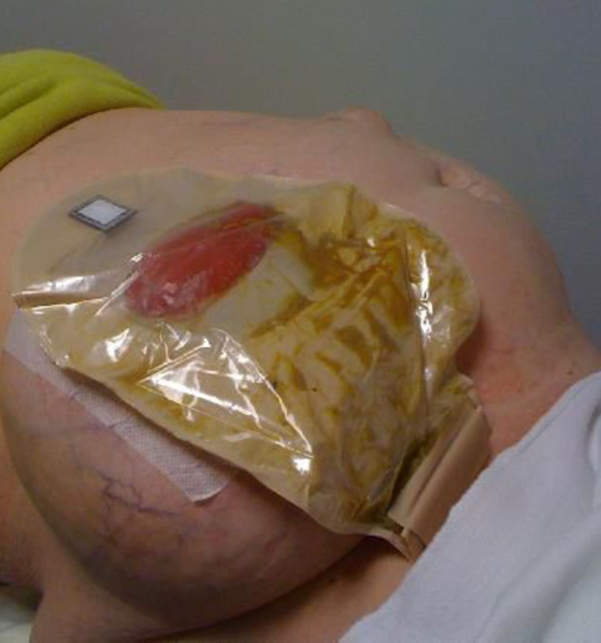 Click for large image | Figure 1. Paracolostomy hernia with a noticeable bulge. |
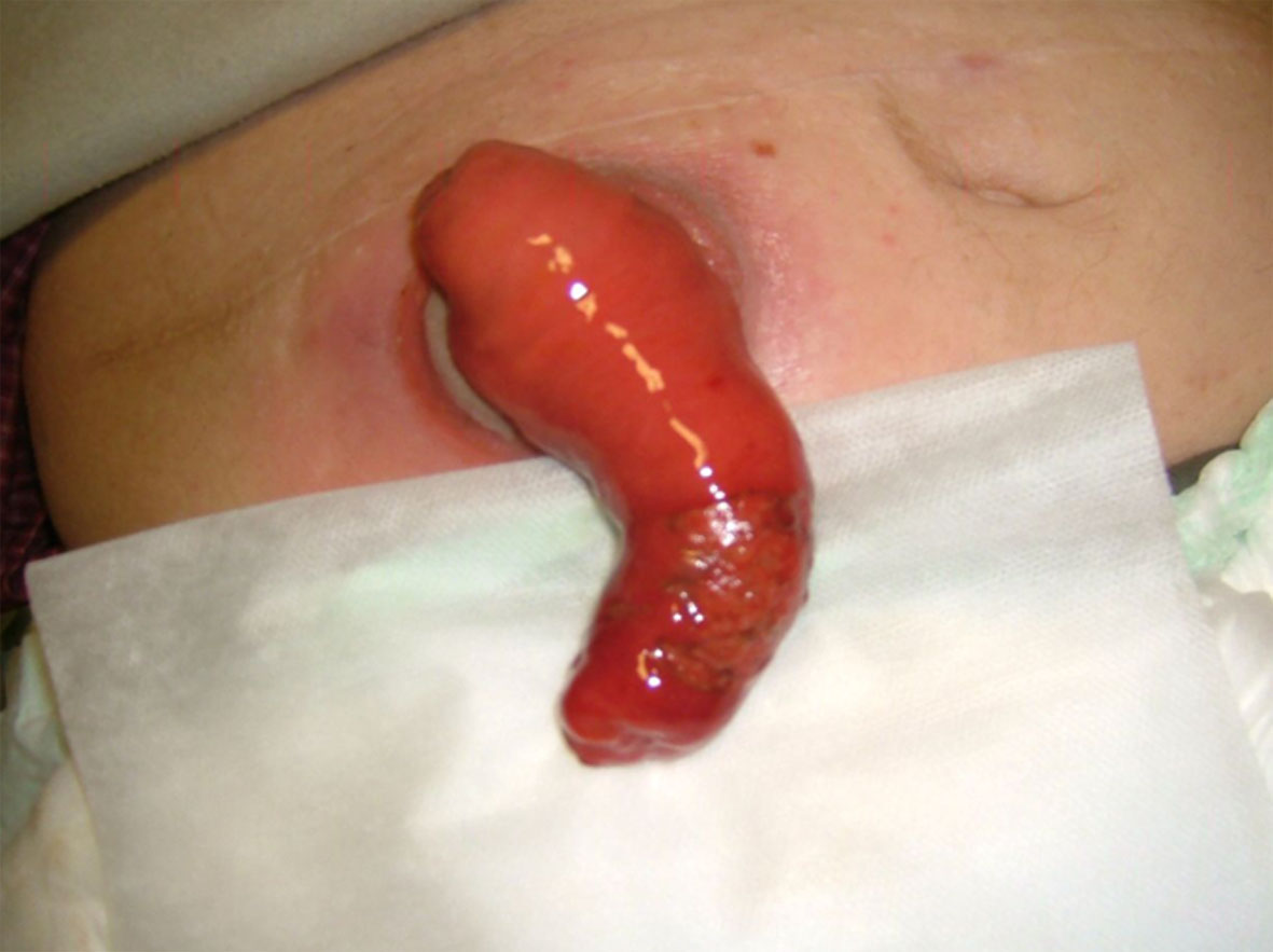 Click for large image | Figure 2. Prolapse loop ileostomy. |
| The Treatment of PH | ▴Top |
Clearly the best treatment for PH is to restore bowel continuity, but this is not always possible. Most of the patients with PH are managed with conservative treatment including diet changes, the use of stomal-supporting devices and medication [10]. About 11-70% of patients require surgery for various PH symptoms [2]. Absolute indications for operation are hernia incarceration, strangulation, obstruction, fistulization, perforation and stomal ischemia. Other indications for surgery are relative and include failure of conservative treatment, pain, ulceration of the surrounding skin and aesthetic reasons. Surgery should be avoided if at all possible in cases of terminal malignant disease or serious co-morbidity conditions such as frailty, severe cardiopulmonary disease, gross obesity and abdominal wall attenuation following multiple previous operations [25]. As in any other elective surgery, a thorough anesthetic evaluation should be done using risk stratification tools such as the American Society of Anesthesiologists (ASA) and Physiological and Operative Severity Score for the enUmeration of Mortality and Morbidity (POSSUM).
| The Surgical Approach for the Repair of PH | ▴Top |
Currently there are a variety of surgical approaches used for the repair of PH. Operations can be divided according to several characteristics: primary site repair versus relocation of stoma, the use of mesh to reinforce the hernia-stoma site or local fascial-aponeurotic repair and also an open versus laparoscopic approach.
The main advantage of primary fascial repair without a mesh is its simplicity which has made it the most prevalent type of repair for many years. An incision is made a few centimeters away from the stoma site followed by a careful dissection and exposure of the hernial sac and orifice. The repair is done using non-absorbable sutures which approximate to the hernial orifice margins. Although simple, in the era of mesh usage this technique has been virtually abandoned due to the high rate of recurrence, estimated at 46-100%, but it is still being used in specific circumstances such as an urgent operation involved with contamination.
Relocation of the stoma as suggested by Goligher [26] has the benefit of using a “fresh” abdominal wall site with the disadvantage of added abdominal dissection. This repair includes dissection of the hernia and stoma along with other parts of the bowel in order to enable stoma repositioning. It can be done through a full laparotomy incision with the risk of celiotomy-related complications or through a near-stoma incision with a nearby new stoma creation [27]. The hernia recurrence rate in this type of repair is lower in comparison to primary fascial repair, but the complication rate is higher along with significantly longer hospitalization. This approach can be difficult in patients with co-morbidities of intra-abdominal adhesions [28].
Although the use of mesh for abdominal wall incisional hernia repair has been known for years, surgeons have tended to avoid using it for repair of PH due to fear of infection. Prosthetic mesh in proximity to the abdominal contents and intestine might also cause other severe complications such as fistulas, adhesions or strictures. Nevertheless, placing a non-absorbable mesh in an attempt to repair a PH was first published in 1977 [29, 30]. Recent studies involving mesh placement during stoma construction suggest that complication rate is low [24]. Currently three types of mesh are being used for PH repair including non-absorbable mesh, double-sided patch and biological meshes, though no conclusive data exist as to which type of mesh is preferable [31].
The mesh repair of PH can be further classified according to its placement within the abdominal wall layer as superficial, retro-muscular and intra-abdominal [2, 32]. Similar to primary fascial repair, superficial PH mesh repair (also termed as onlay repair) involves a remote incision and dissection of the stoma as well as the peritoneal sac and hernial orifice. Closure of the stoma is an important step in order to avoid spillage and contamination. The anchoring of the mesh is done under the subcutaneous layer and above the first aponeurotic-muscle sheet layer which avoids intra-peritoneal mesh placement complications such as adhesions. The stoma is then re-formed [33]. The same basic steps of surgery, meaning adhesiolysis and hernial orifice dissection and peritoneal sac resection are needed in retro-muscular PH mesh repair (also termed as sublay repair) with a major alteration of the positioning of the prosthesis under the muscular layer. For that, a further dissection is carried out in between the abdominal wall layers to enable mesh placement. The peritoneum is then sutured to cover the abdominal content and avoid intra-peritoneal mesh placement complications. A recent meta-analysis has shown that there is no difference in terms of infection, and although not statistically significant, the onlay technique had a higher recurrence rate [32].
The surgical approach to intra-abdominal prosthesis placement differs from other PH repairs by using an old midline or paramedian abdominal incision and avoiding surgery at the contaminated end of the stoma. Adhesions are dissected and the hernial sac content is delivered into the abdominal cavity, and the hernia orifice is then covered with prosthesis. Intra-abdominal mesh repair can be further divided into two subtypes. The Sugarbaker technique which involves laying a mesh patch over the orifice of the hernia leaving the stoma sling laid in between the abdominal wall and the abdominal surface of the mesh [34] and a slit mesh repair through which the bowel can be passed [35].
Laparoscopic PH repair is based on two elementary rules which are complete abdominal wall dissection and the use of surgical mesh for the repair. The dissection includes discovery of the abdominal wall which may necessitate the separation of structures such as the falciform ligament and the pre-vesical space as well as the scar area to allow adequate anchoring and mesh incorporation. The mesh should cover the scar with a broad overlap of at least 5 cm in a similar way to other ventral hernia repairs. Meshes used for laparoscopic approaches must induce strong and rapid incorporation on the parietal side, and they should also prevent adhesions on the visceral side. The mesh might be placed in a modified Sugarbaker technique or slit mesh repair with a keyhole. The modified Sugarbaker technique uses minimally invasive surgery to cover the orifice of the hernia with a mesh similar to the open surgery described earlier [36]. The mesh is then fixed to the fascia with laparoscopic tackers and might be reinforced with sutures according to the surgeon’s preferences. The slit mesh repair might use a designated mesh or surgeon cut mesh with a slit to enable encirclement of the bowel from within the abdomen and a keyhole through which the bowel can be passed (Fig. 3, 4 ) [37]. Different modifications have been suggested including the use of two layers of mesh [38]. According to the literature, recurrence rates are significantly lower with laparoscopic repair using the Sugarbaker compared with the keyhole technique [32].
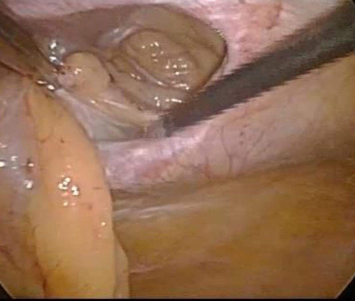 Click for large image | Figure 3. Laparoscopic view of an orrifice of PH. |
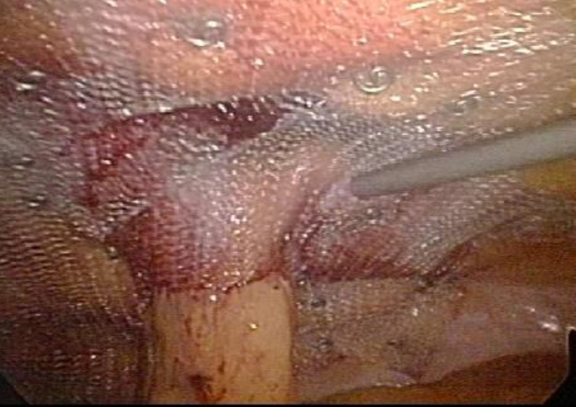 Click for large image | Figure 4. Laparoscopic keyhole repair of PH. |
In summary, the rate of PH is high and influenced by patient-related factors and surgical technique. In spite of the high incidence of PH the appropriate repair technique is not yet defined, and a wide range of surgical options is available. Nevertheless, local repair should be used in special cases such as emergency and frail patient, as the recurrence rates are high. Mesh repair is better than non-mesh repair, and complication rates are low. When performing laparoscopic repair the Sugarbaker technique is preferred over the keyhole technique.
Disclosure
The authors have no financial attachments to disclose.
| References | ▴Top |
- Rosin RD. An obituary to the transverse loop colostomy ('Gone with the wind'). J R Soc Med. 1987;80(12):728-729.
pubmed - Israelsson LA. Parastomal hernias. Surg Clin North Am. 2008;88(1):113-125, ix.
doi pubmed - Kingsnorth A, Leblanc K. Parastomal Hernia. In: Kingsnorth, Andrew, LeBlanc, Karl A. Management of Abdominal Hernias, 3rd ed. London Springer-Verlagon. 2003.
- Smietanski M, Szczepkowski M, Alexandre JA, Berger D, Bury K, Conze J, Hansson B, et al. European Hernia Society classification of parastomal hernias. Hernia. 2014;18(1):1-6.
doi pubmed - Cingi A, Solmaz A, Attaallah W, Aslan A, Aktan AO. Enterostomy closure site hernias: a clinical and ultrasonographic evaluation. Hernia. 2008;12(4):401-405.
doi pubmed - Cingi A, Cakir T, Sever A, Aktan AO. Enterostomy site hernias: a clinical and computerized tomographic evaluation. Dis Colon Rectum. 2006;49(10):1559-1563.
doi pubmed - Mylonakis E, Scarpa M, Barollo M, Yarnoz C, Keighley MR. Life table analysis of hernia following end colostomy construction. Colorectal Dis. 2001;3(5):334-337.
doi pubmed - Kanehisa H, Miyatani M, Azuma K, Kuno S, Fukunaga T. Influences of age and sex on abdominal muscle and subcutaneous fat thickness. Eur J Appl Physiol. 2004;91(5-6):534-537.
doi pubmed - De Raet J, Delvaux G, Haentjens P, Van Nieuwenhove Y. Waist circumference is an independent risk factor for the development of parastomal hernia after permanent colostomy. Dis Colon Rectum. 2008;51(12):1806-1809.
doi pubmed - Arumugam PJ, Bevan L, Macdonald L, Watkins AJ, Morgan AR, Beynon J, Carr ND. A prospective audit of stomas--analysis of risk factors and complications and their management. Colorectal Dis. 2003;5(1):49-52.
doi pubmed - Nastro P, Knowles CH, McGrath A, Heyman B, Porrett TR, Lunniss PJ. Complications of intestinal stomas. Br J Surg. 2010;97(12):1885-1889.
doi pubmed - Pilgrim CH, McIntyre R, Bailey M. Prospective audit of parastomal hernia: prevalence and associated comorbidities. Dis Colon Rectum. 2010;53(1):71-76.
doi pubmed - Carne PW, Robertson GM, Frizelle FA. Parastomal hernia. Br J Surg. 2003;90(7):784-793.
doi pubmed - Williams JG, Etherington R, Hayward MW, Hughes LE. Paraileostomy hernia: a clinical and radiological study. Br J Surg. 1990;77(12):1355-1357.
doi pubmed - Babcock G, Bivins BA, Sachatello CR. Technical complications of ileostomy. South Med J. 1980;73(3):329-331.
doi pubmed - Nguyen MH, Pittas F. How large should a skin trephine be for an end stoma? Aust N Z J Surg. 1999;69(9):675-676.
doi - Koltun L, Benyamin N, Sayfan J. Abdominal stoma fashioned by a used circular stapler. Dig Surg. 2000;17(2):118-119.
doi pubmed - Sjodahl R, Anderberg B, Bolin T. Parastomal hernia in relation to site of the abdominal stoma. Br J Surg. 1988;75(4):339-341.
doi pubmed - Londono-Schimmer EE, Leong AP, Phillips RK. Life table analysis of stomal complications following colostomy. Dis Colon Rectum. 1994;37(9):916-920.
doi pubmed - Leong AP, Londono-Schimmer EE, Phillips RK. Life-table analysis of stomal complications following ileostomy. Br J Surg. 1994;81(5):727-729.
doi pubmed - Porter JA, Salvati EP, Rubin RJ, Eisenstat TE. Complications of colostomies. Dis Colon Rectum. 1989;32(4):299-303.
doi pubmed - Israelsson LA. Preventing and treating parastomal hernia. World J Surg. 2005;29(8):1086-1089.
doi pubmed - Janes A, Cengiz Y, Israelsson LA. Randomized clinical trial of the use of a prosthetic mesh to prevent parastomal hernia. Br J Surg. 2004;91(3):280-282.
doi pubmed - Hansson BM. Parastomal hernia: treatment and prevention 2013; where do we go from here? Colorectal Dis. 2013;15(12):1467-1470.
doi pubmed - Tadeo-Ruiz G, Picazo-Yeste JS, Moreno-Sanz C, Herrero-Bogajo ML. [Parastomal hernias: background, current status and future prospects]. Cir Esp. 2010;87(6):339-349.
doi pubmed - Goligher JC. Intestinal Stoma. Goligher JC Surgery of the Anus, Rectum and Colon. 5th revised edition. London Bailliere Tindall; 1984.
- Rubin MS, Schoetz DJ, Jr., Matthews JB. Parastomal hernia. Is stoma relocation superior to fascial repair? Arch Surg. 1994;129(4):413-418; discussion 418-419.
doi pubmed - Baig MK, Larach JA, Chang S, Long C, Weiss EG, Nogueras JJ, Wexner SD. Outcome of parastomal hernia repair with and without midline laparotomy. Tech Coloproctol. 2006;10(4):282-286.
doi pubmed - Rosin JD, Bonardi RA. Paracolostomy hernia repair with Marlex mesh: a new technique. Dis Colon Rectum. 1977;20(4):299-302.
doi - Morris-Stiff G, Hughes LE. The continuing challenge of parastomal hernia: failure of a novel polypropylene mesh repair. Ann R Coll Surg Engl. 1998;80(3):184-187.
pubmed - Sheen AJ. Prosthetics in hernia repair. Surg Today. 2005;35(3):196-198.
doi pubmed - Hansson BM, Slater NJ, van der Velden AS, Groenewoud HM, Buyne OR, de Hingh IH, Bleichrodt RP. Surgical techniques for parastomal hernia repair: a systematic review of the literature. Ann Surg. 2012;255(4):685-695.
doi pubmed - Tekkis PP, Kocher HM, Payne JG. Parastomal hernia repair: modified thorlakson technique, reinforced by polypropylene mesh. Dis Colon Rectum. 1999;42(11):1505-1508.
doi pubmed - Sugarbaker PH. Peritoneal approach to prosthetic mesh repair of paraostomy hernias. Ann Surg. 1985;201(3):344-346.
doi pubmed - Hansson BM, van Nieuwenhoven EJ, Bleichrodt RP. Promising new technique in the repair of parastomal hernia. Surg Endosc. 2003;17(11):1789-1791.
doi pubmed - Pastor DM, Pauli EM, Koltun WA, Haluck RS, Shope TR, Poritz LS. Parastomal hernia repair: a single center experience. JSLS. 2009;13(2):170-175.
pubmed - Mizrahi H, Bhattacharya P, Parker MC. Laparoscopic slit mesh repair of parastomal hernia using a designated mesh: long-term results. Surg Endosc. 2012;26(1):267-270.
doi pubmed - Berger D, Bientzle M. Laparoscopic repair of parastomal hernias: a single surgeon's experience in 66 patients. Dis Colon Rectum. 2007;50(10):1668-1673.
doi pubmed
This is an open-access article distributed under the terms of the Creative Commons Attribution License, which permits unrestricted use, distribution, and reproduction in any medium, provided the original work is properly cited.
Journal of Current Surgery is published by Elmer Press Inc.