Journal of Current Surgery, ISSN 1927-1298 print, 1927-1301 online, Open Access
Article copyright, the authors; Journal compilation copyright, J Curr Surg and Elmer Press Inc
Journal website http://www.currentsurgery.org

Original Article

Volume 5, Number 4, December 2015, pages 183-187


Comparative Analysis of Anatomy Following Intraoperative Cholangiograms in Elective Laparoscopic Cholecystectomies

Figures

Figure 1.
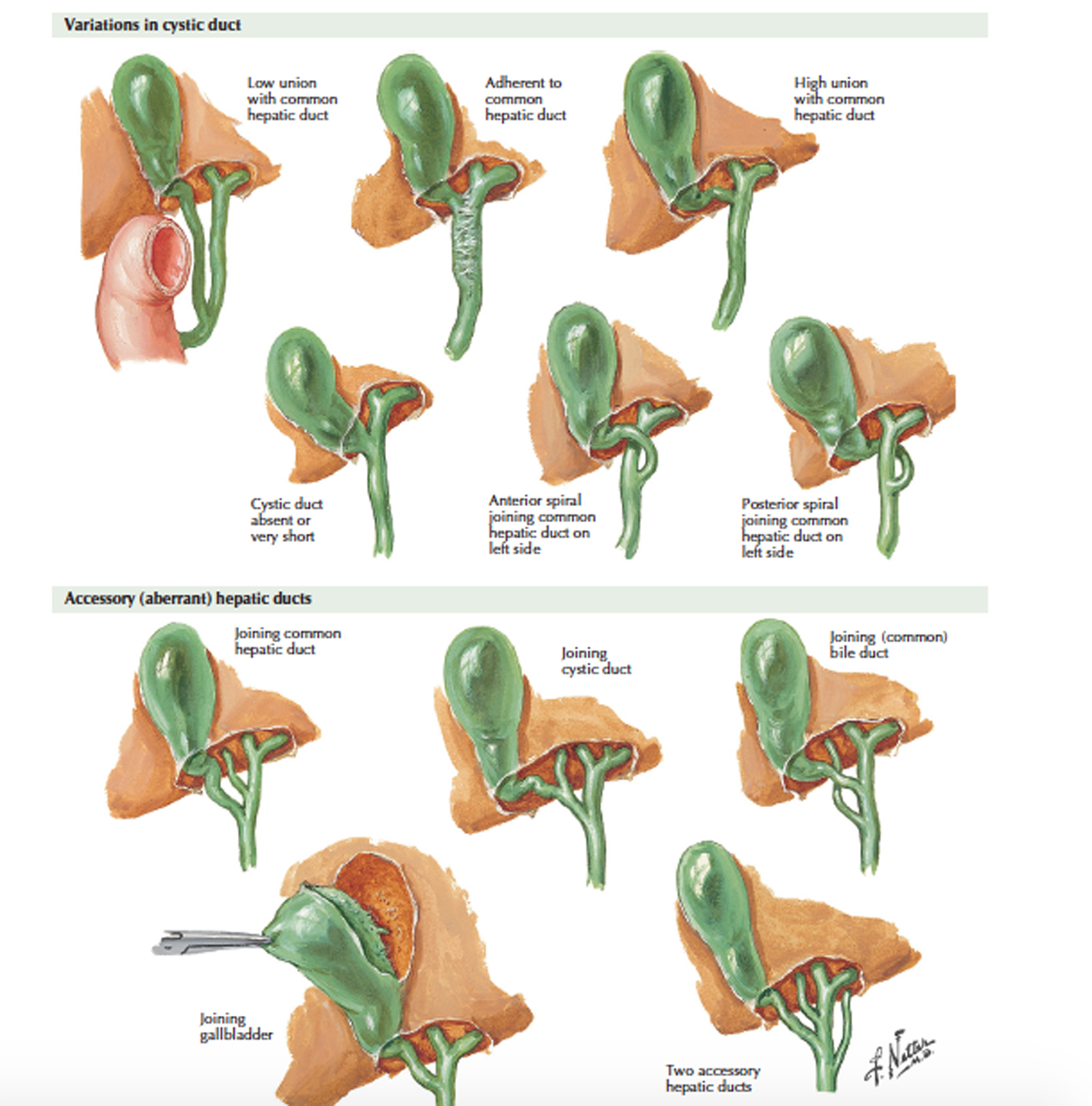
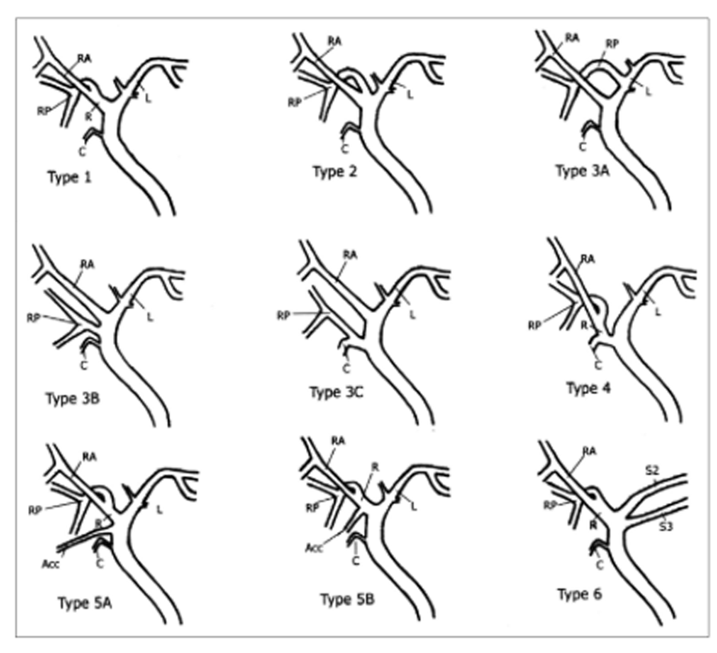
Figure 1. Schematic drawing of IHD anatomy. Type 1 is typical. Type 2 involves triple confluence, the simultaneous emptying of the RASD, RPSD and LHD into the CHD. In type 3, the RPSD drains anomalously, and in type 4, the RHD drains into the cystic duct. In type 5, an accessory duct is present, and in type 6, segments II and III drain individually into the RHD or CHD. Type 7 shows unclassified or complex variation. R: right hepatic duct; L: left hepatic duct; RA: right anterior segmental duct; RP: right posterior segmental duct; C: cystic duct; Acc: accessory duct [7].
Figure 2.
Figure 2. Variations in hepatic and cystic duct [1].
Figure 3.
Figure 3. Intraoperative cholangiogram showing normal ductal anatomy.
Figure 4.
Figure 4. RPSD draining into CHD.
Figure 5.
Figure 5. Triple confluence of RASD, RPSD, and LHD into CHD.

Tables

Table 1. Number and Type of Anatomic Variants
 
AnatomyNumber of patients
Normal anatomy12
Variants
  Accessory hepatic duct draining into CHD2
  RPSD draining into CHD2
  Triple confluence of RASD, RPSD and left hepatic duct into CHD2
  RPSD draining into LHD1


Table 2. Complications Associated With Variants
 
AnatomyIncreased intraoperative hemorrhageCBD injuryOther ductal injuries
Normal1*00
Variants in anatomy
  Triple confluence of RASD, RPSD, and LHD into CHD000
  Accessory hepatic duct draining into CHD000
  RPSD draining into CHD000
  RPSD draining into LHD000