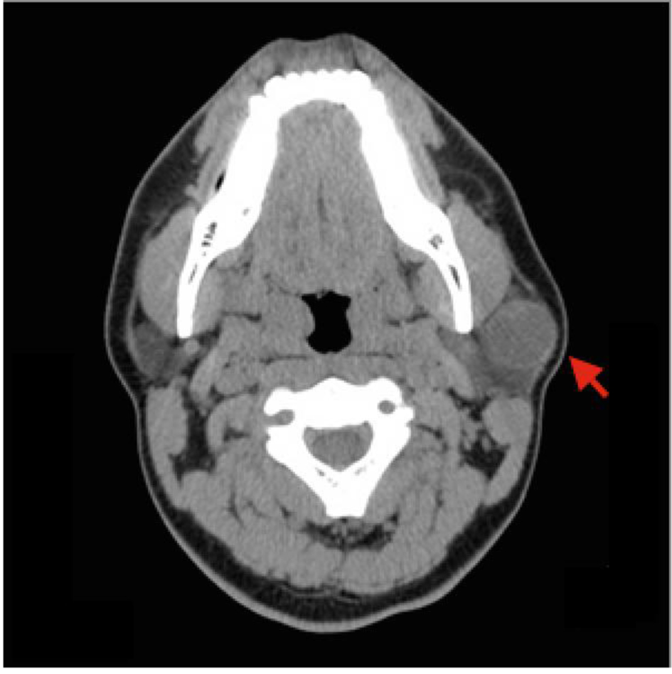
Figure 1. Computed tomography (CT) of the neck. Volumetric non-contrast CT of the soft tissues of the neck demonstrates a well-circumscribed 2.2 × 2.2 × 2.1 cm cystic tumor in the tail of the left parotid gland (red arrow).
| Journal of Current Surgery, ISSN 1927-1298 print, 1927-1301 online, Open Access |
| Article copyright, the authors; Journal compilation copyright, J Curr Surg and Elmer Press Inc |
| Journal website http://www.currentsurgery.org |
Case Report
Volume 6, Number 1, March 2016, pages 30-32
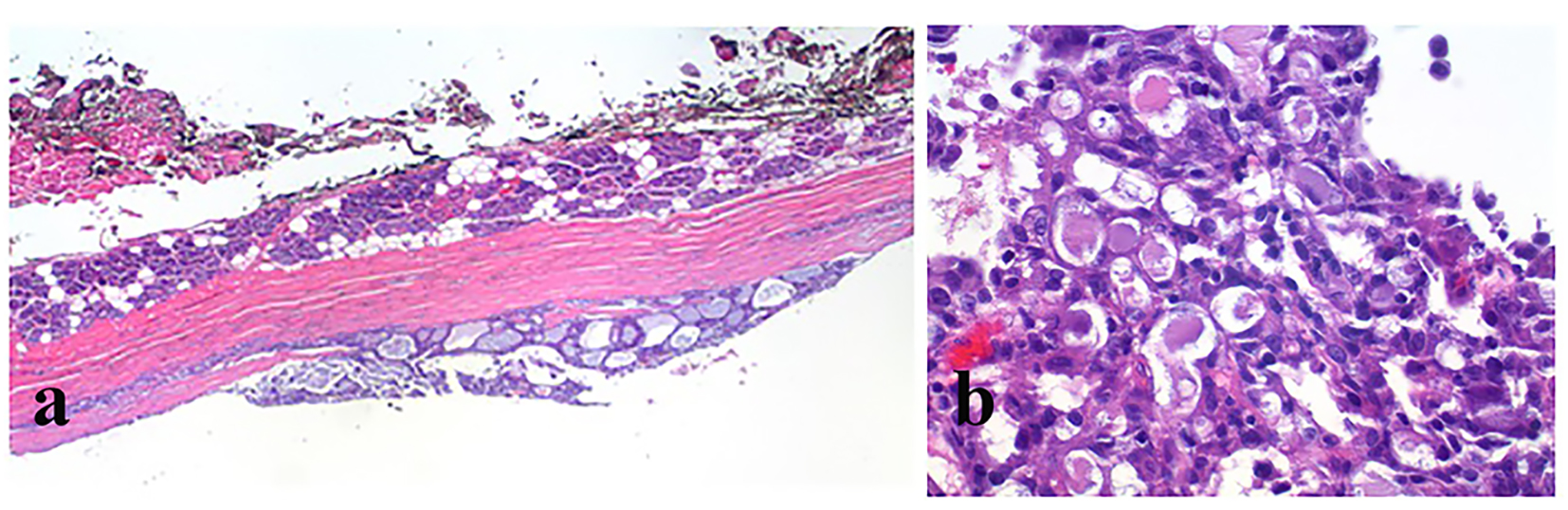
Surgical Excision of a Rare Case of Mammary Analogue Secretory Carcinoma: A Case Review
Figures