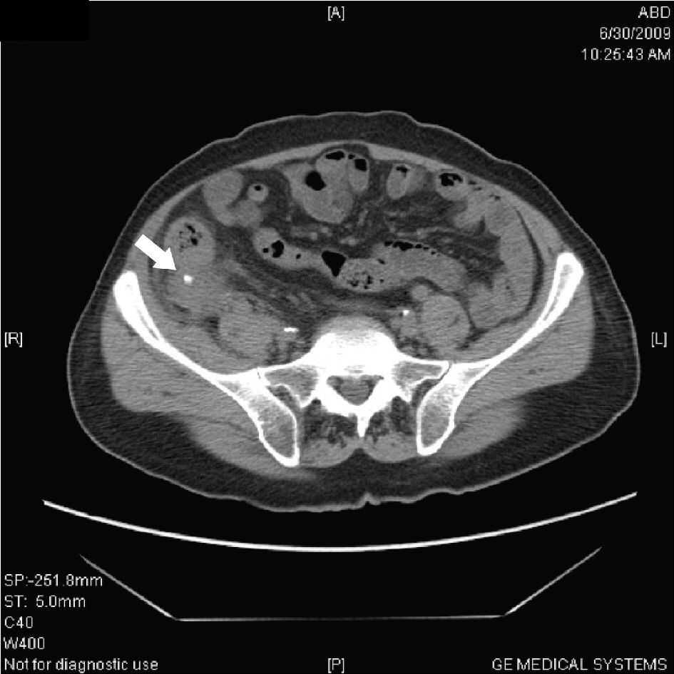
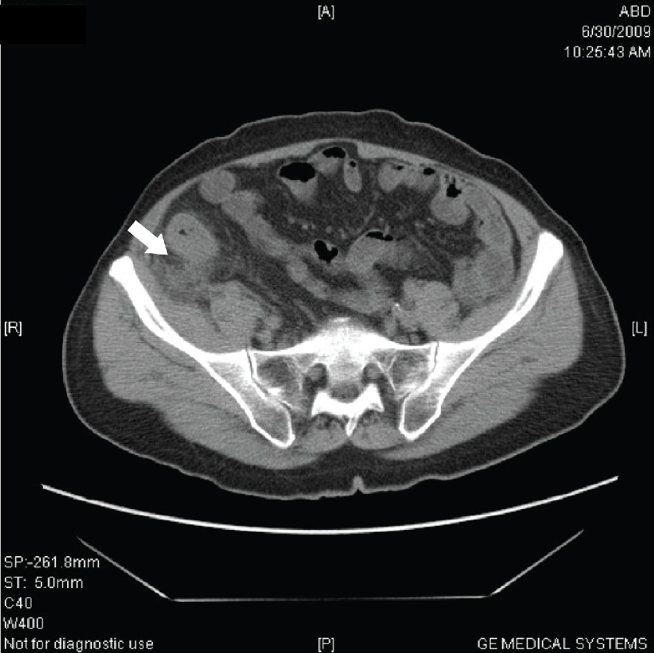
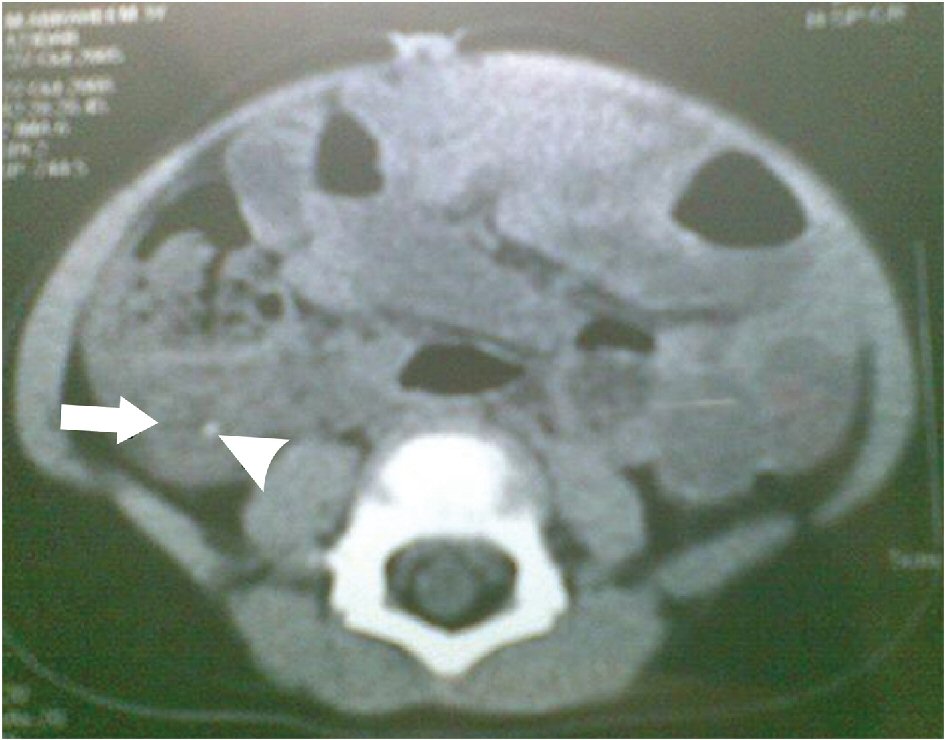
Figure 1. FACT scan demonstrating appendicolith.
| Journal of Current Surgery, ISSN 1927-1298 print, 1927-1301 online, Open Access |
| Article copyright, the authors; Journal compilation copyright, J Curr Surg and Elmer Press Inc |
| Journal website http://www.currentsurgery.org |
Original Article
Volume 8, Number 1-2, June 2018, pages 7-12
Focused Abdominal Computed Tomography in Clinically Suspected Adolescent Acute Appendicitis
Figures
Tables
| Number of patients (N) | Percentage (%) | |
|---|---|---|
| Diagnosed on FACT scan | 85 | 89.4 |
| Not diagnosed on FACT scan | 10 | 10.5 |
| Total | 95 | 100 |
| Number of patients (N) | Percentage (%) | |
|---|---|---|
| Yes | 88 | 92.6 |
| No | 7 | 7.4 |
| Total | 95 | 100 |
| Sensitivity | 97.32% |
| Specificity | 88.42% |
| Positive predictive value | 98.8% |
| Negative predictive value | 80.0% |
| Diagnostic accuracy | 96.8% |