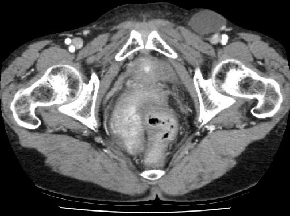
Figure 1. Computed tomography (CT) shows a cystic mass (3.6 × 5.6 cm) at left inguinal region (previous size: 3.6 × 5.1 cm).
| Journal of Current Surgery, ISSN 1927-1298 print, 1927-1301 online, Open Access |
| Article copyright, the authors; Journal compilation copyright, J Curr Surg and Elmer Press Inc |
| Journal website http://www.currentsurgery.org |
Case Report
Volume 8, Number 3-4, November 2018, pages 38-40
A Mesothelial Cyst Presenting as Inguinal Mass: Two Case Reports and Literature Review
Figures
Table
| Authors (year) | Sex/age | Misdiagnosis | Location | Size(cm) | Reducibility | Hernia sac |
|---|---|---|---|---|---|---|
| M: male; F: female. | ||||||
| Aarabi et al (2010) [9] | M/10 | Inguinal mass | Spermatic cord | 6.5 × 1.8 | Yes | Yes |
| Kim et al (2010) [10] | F/76 | Mimicking a metastasis | Round ligament | 21 × 10 | No | No |
| Vaos et al (2009) [11] | M/2 | Undescended testis | Spermatic cord | 1.5 × 1.0 | No | No |
| Ryley et al (2004) [2] | F/31 | Inguinal hernia | Round ligament | 4.8 × 2.3 | No | N0 |