Journal of Current Surgery, ISSN 1927-1298 print, 1927-1301 online, Open Access
Article copyright, the authors; Journal compilation copyright, J Curr Surg and Elmer Press Inc
Journal website http://www.currentsurgery.org

Case Report

Volume 8, Number 3-4, November 2018, pages 41-43


Abdominal Wall Mass: An Unusual Complication of a Ventral Hernia Repair

Figures

Figure 1.
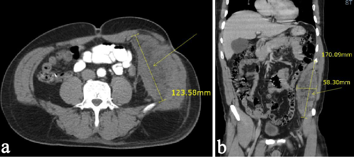
Figure 1. (a, b) CT scan of the abdomen and pelvis demonstrating an 11-cm long extraperitoneal soft tissue density along the left hemi-abdomen.
Figure 2.
Figure 2. (a, b) CT scan of the abdomen and pelvis after CT-guided biopsy demonstrating a mass increased in size of 17 × 5.8 cm with a slight increase in the hemorrhage associated with the left flank/ abdominal wall mass.
Figure 3.
Figure 3. (a, b) MRI of the abdomen and pelvis demonstrating a hemorrhage associated in the left lateral abdominal wall mass. The hyperintensity shown on the MRI marked by the arrowhead indicates the presence of a hemorrhage.
Figure 4.
Figure 4. (a, b) Abdominal wall mass originating from hernia repair incision scar.