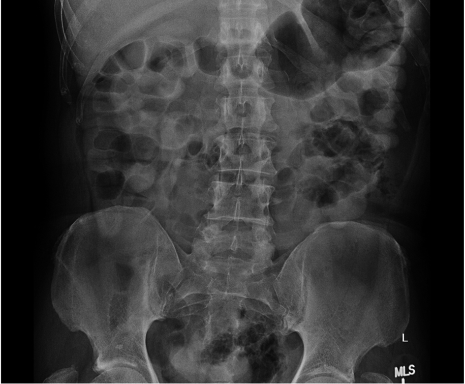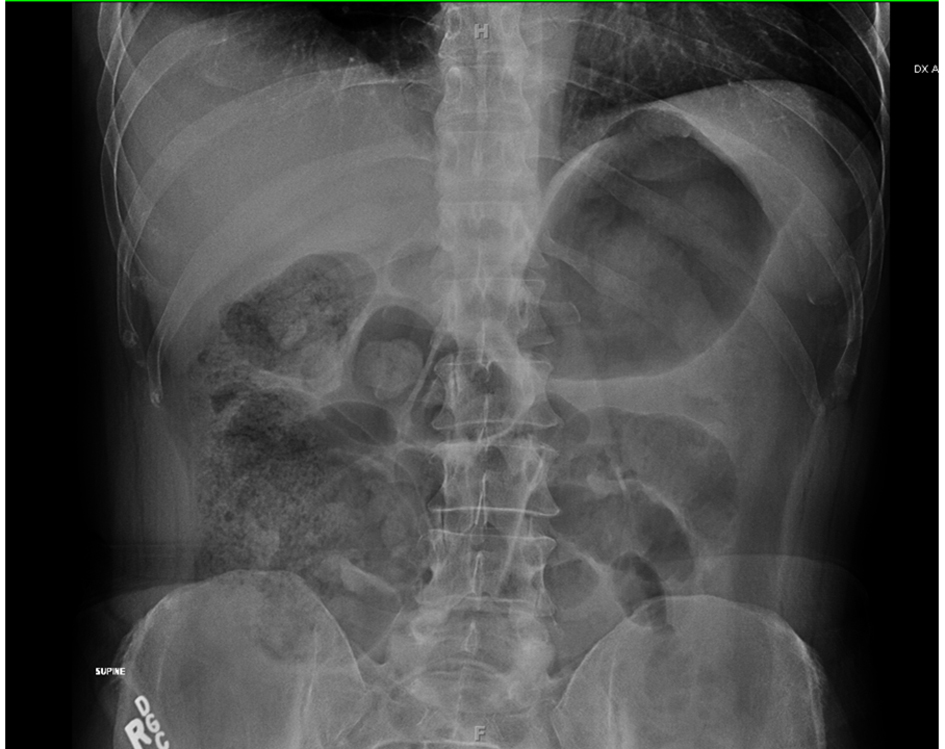
Figure 1. Antero-posterior plain abdominal X ray at presentation, showing distension of small bowel loops with air fluid levels.
| Journal of Current Surgery, ISSN 1927-1298 print, 1927-1301 online, Open Access |
| Article copyright, the authors; Journal compilation copyright, J Curr Surg and Elmer Press Inc |
| Journal website http://www.currentsurgery.org |
Case Report
Volume 2, Number 3, June 2012, pages 110-112
Spontaneous Resolution of Primary Small Bowel Volvulus With Oral Contrast
Figures