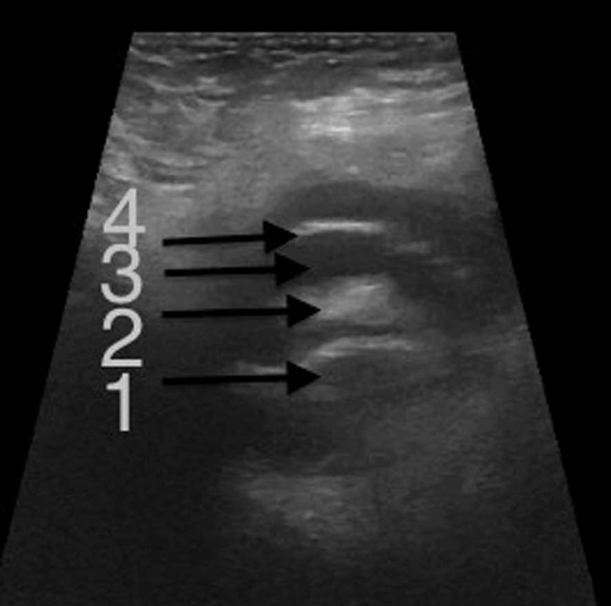
Figure 1. Transverse scan ( sonography) of the right groin showing the fluid-filled hernia sac containing the appendix. 1 = appendicitis; 2 = mesoappendix; 3 = effusion; 4= hernia sac.
| Journal of Current Surgery, ISSN 1927-1298 print, 1927-1301 online, Open Access |
| Article copyright, the authors; Journal compilation copyright, J Curr Surg and Elmer Press Inc |
| Journal website http://www.currentsurgery.org |
Case Report
Volume 3, Number 2, October 2013, pages 92-94
Management of the Aymand’s Hernia in Laparoscopy
Figures