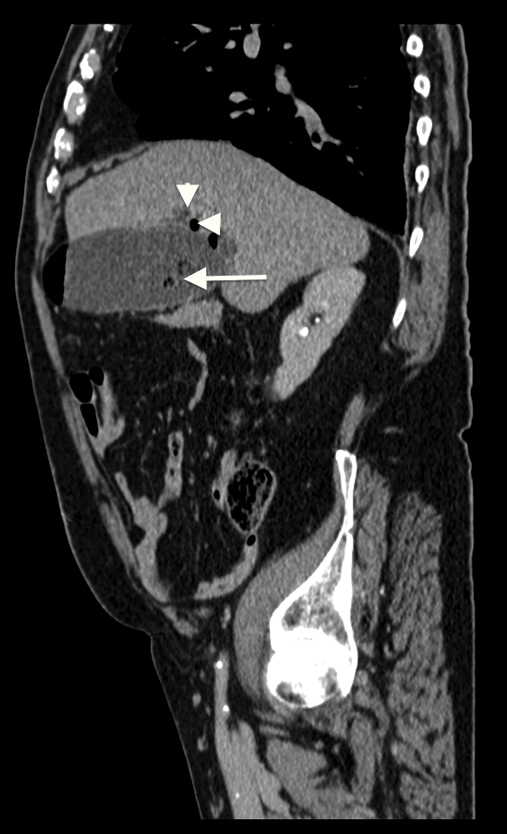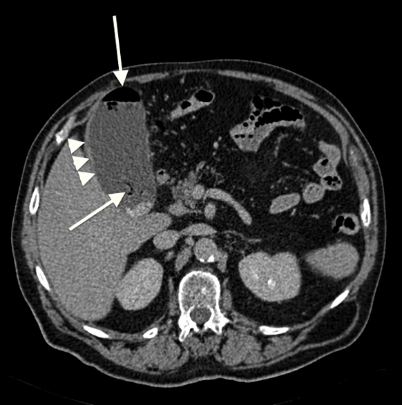
Figure 1. CT scan (axial axis) shows air in the gall bladder lumen with an air-fluid level (arrow) and gall bladder wall thickening (arrowhead).
| Journal of Current Surgery, ISSN 1927-1298 print, 1927-1301 online, Open Access |
| Article copyright, the authors; Journal compilation copyright, J Curr Surg and Elmer Press Inc |
| Journal website http://www.currentsurgery.org |
Case Report
Volume 4, Number 1, February 2014, pages 31-33
Acute Emphasematous Cholecystitis
Figures