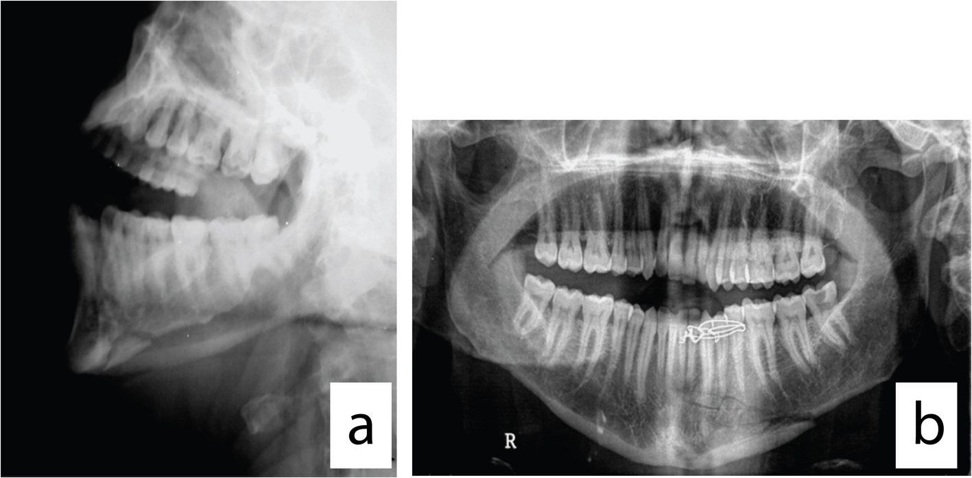
Figure 1. (a) Mandibular lateral oblique X-ray view for evaluation of the comminuted fracture. (b) Mandibular panoramic X-ray view for evaluation of the comminuted fracture.
| Journal of Current Surgery, ISSN 1927-1298 print, 1927-1301 online, Open Access |
| Article copyright, the authors; Journal compilation copyright, J Curr Surg and Elmer Press Inc |
| Journal website http://www.currentsurgery.org |
Original Article
Volume 4, Number 3, September 2014, pages 86-90
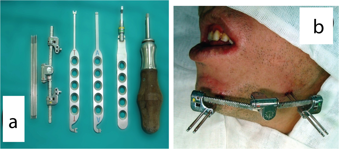
Use of Extraoral Multidirectional Distractor as an External Pin Fixator: A Novel Technique in the Management of Comminuted Mandibular Fracture
Figures