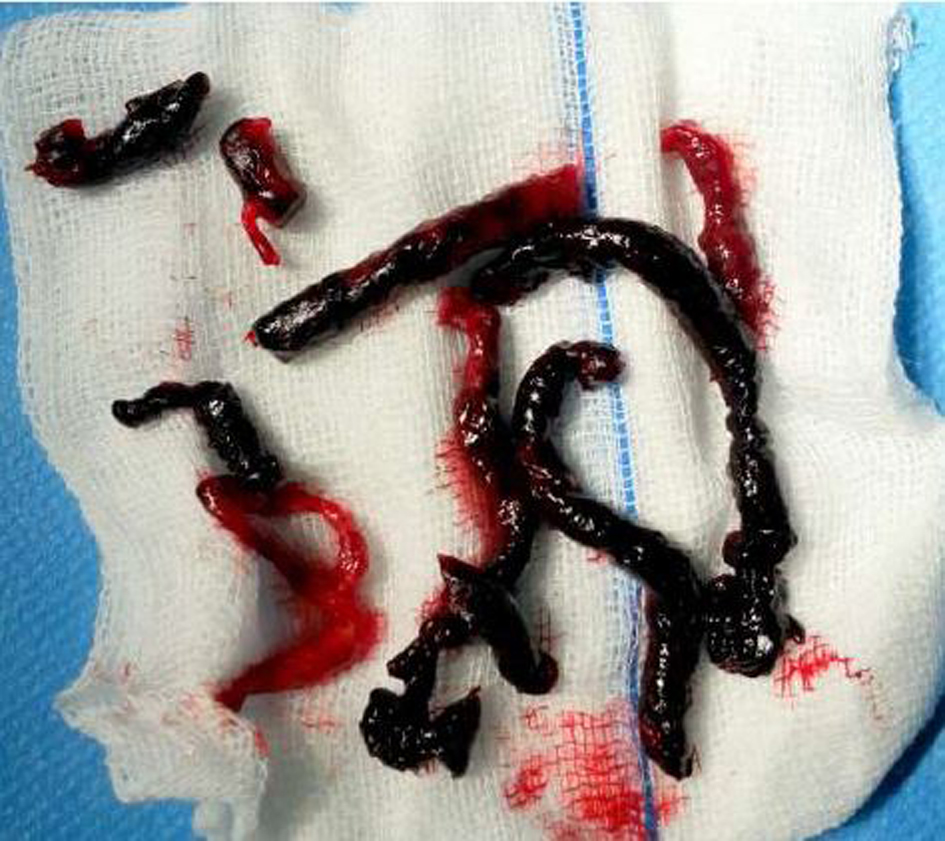
Figure 1. Clots taken out: post-embolectomy.
| Journal of Current Surgery, ISSN 1927-1298 print, 1927-1301 online, Open Access |
| Article copyright, the authors; Journal compilation copyright, J Curr Surg and Elmer Press Inc |
| Journal website http://www.currentsurgery.org |
Case Report
Volume 5, Number 4, December 2015, pages 209-212
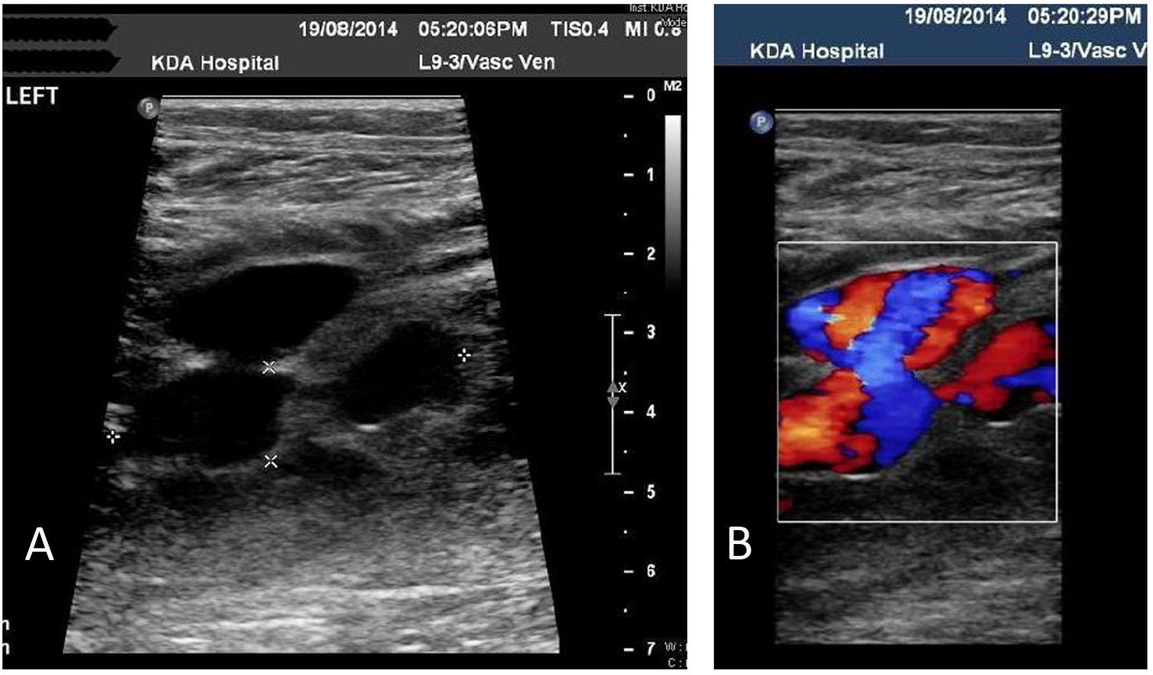
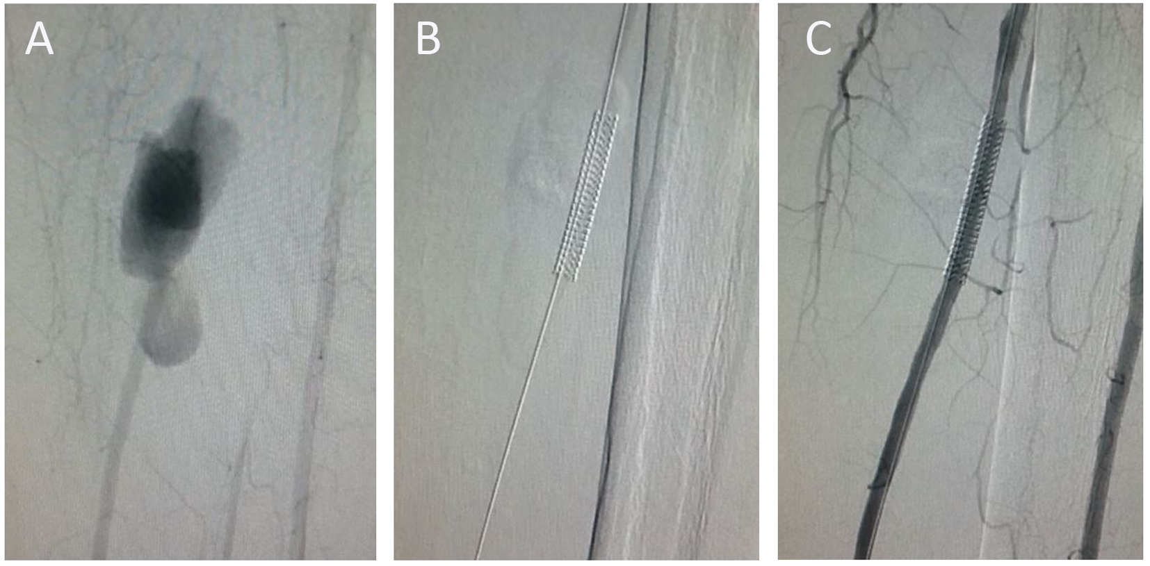
Pseudoaneurysm of Posterior Tibial Artery Management: Case Report and Review of Literature
Figures