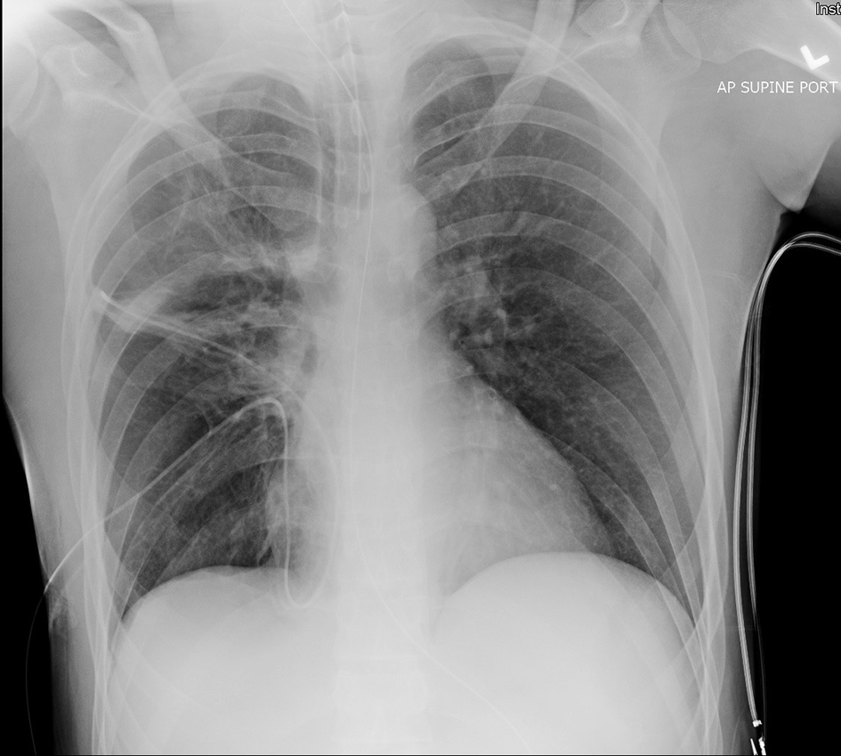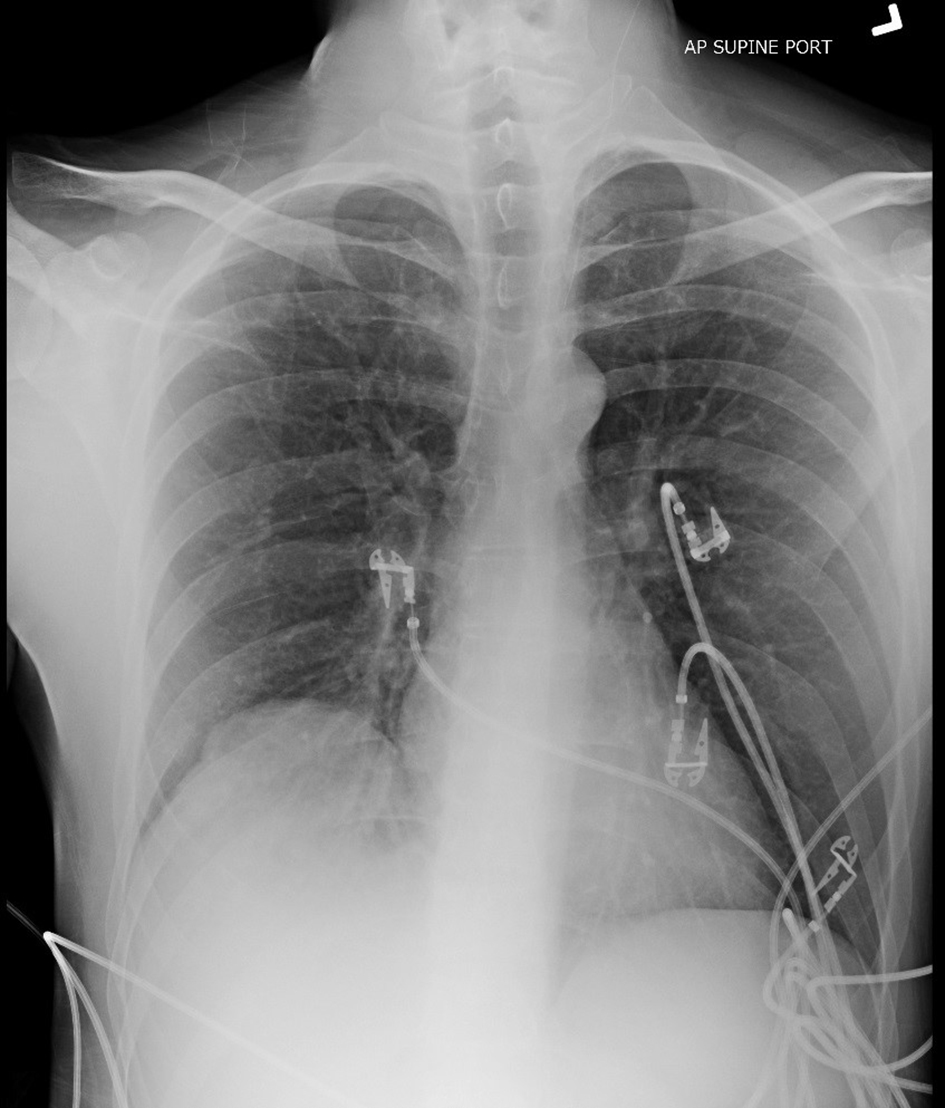
Figure 1. Pre-operative portable AP supine chest X-ray demonstrating elevated right hemidiaphragm.
| Journal of Current Surgery, ISSN 1927-1298 print, 1927-1301 online, Open Access |
| Article copyright, the authors; Journal compilation copyright, J Curr Surg and Elmer Press Inc |
| Journal website http://www.currentsurgery.org |
Case Report
Volume 6, Number 1, March 2016, pages 33-36
Diagnostic Laparoscopy for Right Hemidiaphragm Rupture With Laparotomy Repair: A Case Report
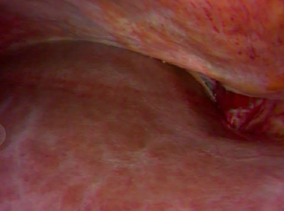
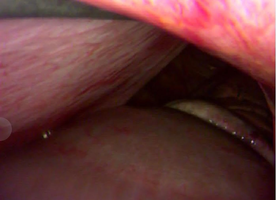
Figures