Journal of Current Surgery, ISSN 1927-1298 print, 1927-1301 online, Open Access
Article copyright, the authors; Journal compilation copyright, J Curr Surg and Elmer Press Inc
Journal website http://www.currentsurgery.org

Original Article

Volume 7, Number 3, September 2017, pages 27-34


Causes and Treatment of Varicose Recurrence in the Popliteal Region

Figures

Figure 1.
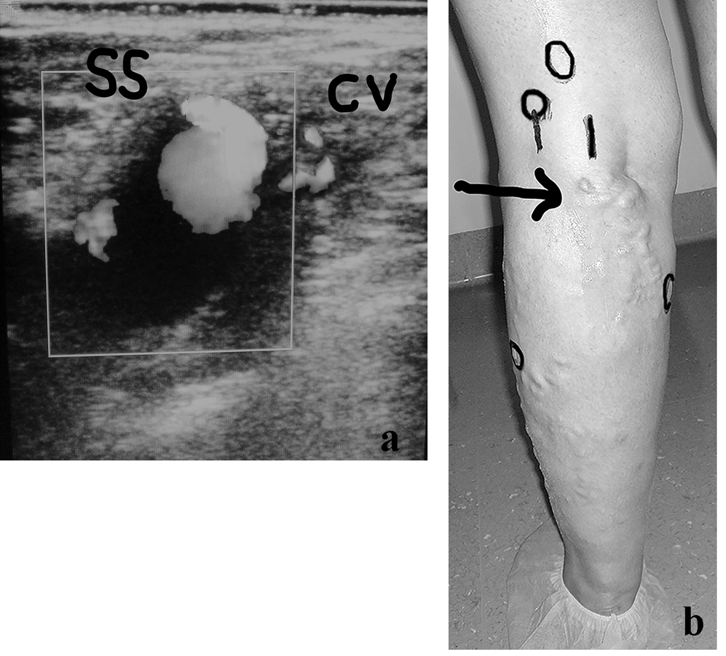
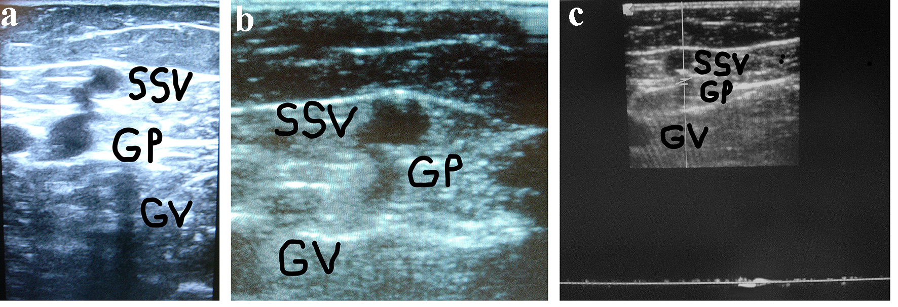
Figure 1. (a) A woman of 67 years of age. Appearance of symptomatic VR in the PR 4 years after the previous surgical procedure for varicose veins at the PR. Cross scanning of DUS examination at the PR: incompetent gastrocnemius perforator (GP) connected with the long residual SSV stump. The internal gastrocnemius vein (GV) is visible. (b) Treatment: SPJ surgical interruption and intraoperative DUS-guided foam sclerotherapy of gastrocnemius perforator (GP) and residual SSV. DUS examination after 30 days: the SS stump is not compressible and partially hyper-echogenic from early fibrosis (SSV), the gastrocnemius perforator (GP) is hyper-echogenic from early complete fibrosis in the subfascial segment, the internal gastrocnemius vein (GV) is patent and compressible. (c) DUS examination after 30 days: the gastrocnemius perforator (GP) is occluded with no flow into the lumen, the SS stump is not compressible and hyper-echogenic from advanced fibrosis (SSV), the internal gastrocnemius vein (GV) is patent and compressible.
Figure 2.
Figure 2. (a) A woman of 42 years of age, operated on for VVLL at the SPJ. Appearance of symptomatic VR in the PR, belonging to a residual SSV, after 2 years. The clinical and preliminary DUS examination demonstrated an aneurysm of the proximal long SSV incompetent stump (SS) connected with a small incompetent collateral vein (CV). (b) Preoperative skin mapping. A more extended DUS examination detected the following combined sources of reflux which are visible in the preoperative skin drawing: aneurysm of the residual SS, double high non-saphenous popliteal perforator, connection with the medial accessory of the GSV as main causes of VR at the PR; low gastrocnemius and peroneal perforators. The black arrow indicates the SS aneurysm. The black circles correspond to the four incompetent perforators.

Tables

Table 1. Distribution of VR at the Junctions in 1,831 Limbs of 1,760 Patients
 
Site of varicose recurrenceN%
SFJ: sapheno-femoral junction; PR: popliteal region. Causistry of the years 2005 - 2016.
Limbs1,831100
Varicose recurrence SFJ - SPR20711.3
Varicose recurrence PR (isolated)19
19/207
1.0
9.1


Table 2. Characteristics of 19 Limbs of 17 Patients Affected With Varicose Recurrence (VR) at the PR
 
MeanMin.Max.
C2-C6: CEAP clinical classification; CHIVA: cure hemodinamique insuffisance veineuse ambulatoire; EVLA : endovenous laser ablation.
Patients (17)
Years of age38.82773
Years from surgery and VR4.8112
N%
Patients17100
  Males741.1
  Females1058.8
Symptomatic (C2-C6)1694.1
Limbs (bilateral 2)19100
  Right736.8
  Left1263.1
  C21263.1
  C3210.5
  C4210.5
  C5210.5
  C615.2
Previous treatments
  Surg. interruption + stripping842.1
  Low ligation +/- stripping SSV631.5
  CHIVA315.7
  EVLA210.5


Table 3. Treatment Performed in 19 Limbs of 17 Patients (Two Bilateral) Affected With VR at the PR
 
TreatmentN%
DUS: duplex ultrasound examination.
Surgical revision interruption + sclerotherapy1368.4
Surgical revision interruption + short SSV stripping15.2
USD-guided sclerotherapy421.0
Conservative treatment15.2
Postoperative sclerotherapy526.3
Elastic compression19100


Table 4. Residual Varicose Veins and Anatomical Findings Detected by DUS Examination in 19 Limbs and by Direct Surgical Dissection in 14
 
Sites of VRN%
Comparison DUS/surgery analyzed by exact Fisher’s test (P = 1). SSV: short saphenous vein; GSV: great saphenous vein; VV: varicose vein. Sapheno-politeal junction: normal outflow of the SSV. Popliteal region: absence of the SSV outflow. In cursive writing 1 aneurysmatic of the 13 saphenous stumps. Percentages calculated on the total of 19 limbs.
Sapheno-popliteal junction1473.6
  Saphenous stump +/- residual tributary (1 aneurysmatic = 5.2%)1368.4
  SSV persistence in a previous duplicity210.5
  Popliteal perforator (non saphenous) (combined)210.5
  Medial gastrocnemius perforator210.5
  Medial communicating SSV-GSV315.7
  Medial accessory (Giacomini)15.2
  Gluteal perforator15.2
  Lateral perforator of the thigh15.2
  VV of the sciatic nerve00
  Neovascularization by neoangiogenesis (suspected)00
  Complex collateral circulation (cavernoma)421.0
Sapheno-popliteal region526.3
  Saphenous Stump - outflow into superficial femoral15.2
  SSV outflowing into the medial accessory (Giacomini)15.2
  Popliteal perforator (non saphenous)210.5
  Medial gastrocnemius perforator210.5
  VV of the sciatic nerve00
  Neovascularization by neoangiogenesis00
  Complex collateral circulation (cavernoma)00