Journal of Current Surgery, ISSN 1927-1298 print, 1927-1301 online, Open Access
Article copyright, the authors; Journal compilation copyright, J Curr Surg and Elmer Press Inc
Journal website http://www.currentsurgery.org

Case Report

Volume 7, Number 4, December 2017, pages 64-68


Intimal Angiosarcoma of the Aorta: Single Case Report and Effective Surgical Management

Figures

Figure 1.
Figure 1. CT and HRCT. (a) CTA 3D imaging of aorta pre-operatively. (b) HRCT descending aorta pre-operatively. (c) HRCT aortic arch pre-operatively. (d) CTA 3D imaging of aorta post-operatively. (e) HRCT descending aorta post-operatively. (f) HRCT aortic arch post-operatively.
Figure 2.
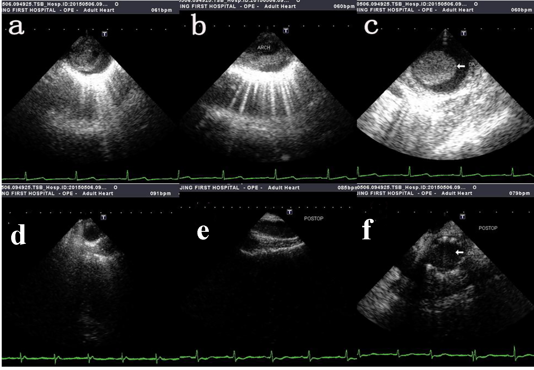
Figure 2. Intra-operative transesophageal echocardiography showing tumor mass protruding into the lumen. (a, b, c) The pre-operative transesophageal echocardiography of ascending aorta, aortic arch and thoracic descending aorta respectively. (d, e, f) The post-operative transesophageal echocardiography of ascending aorta, aortic arch and thoracic descending aorta respectively.
Figure 3.
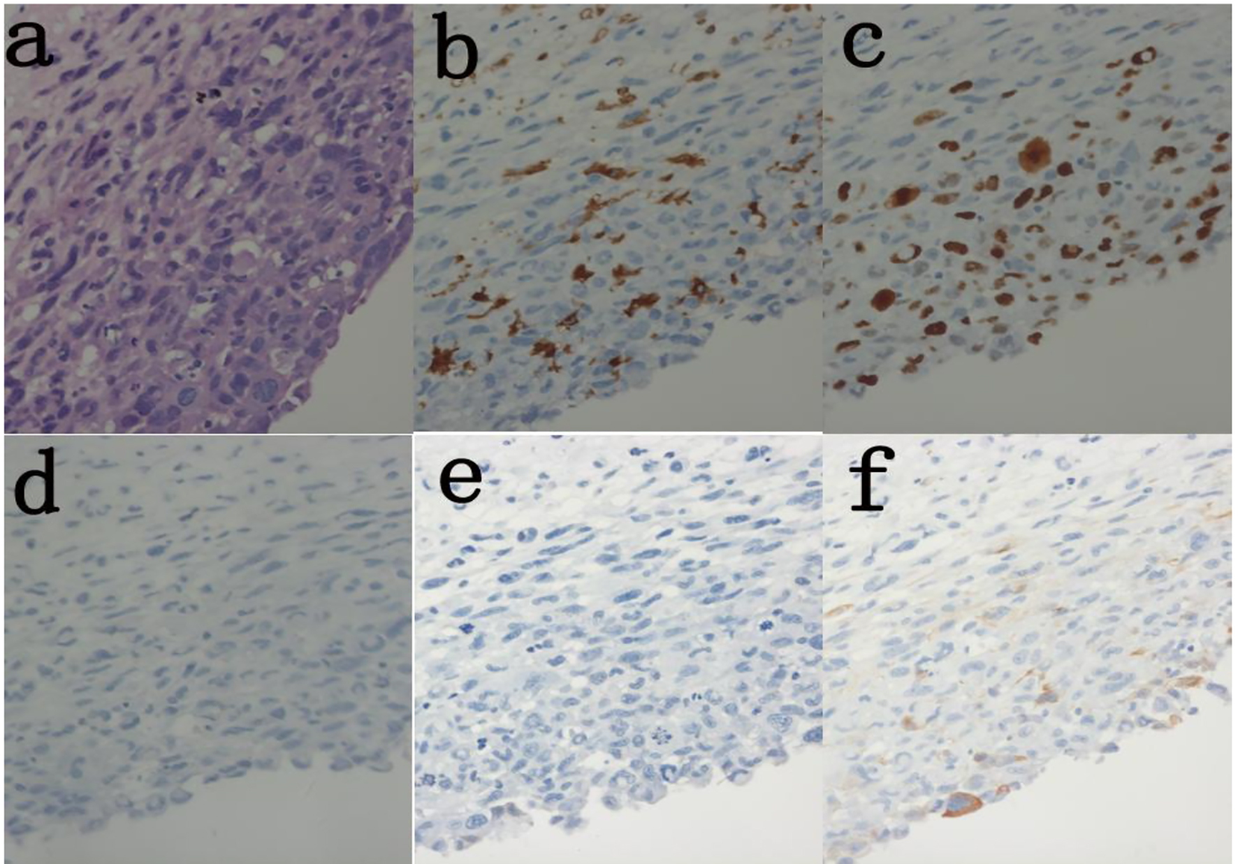
Figure 3. Histolopathology of excised aortic wall lesion shows malignant infiltration of smooth muscle cells. (a) H&E (× 400). (b) Immunoassay Ki-67 (30%) (× 400). (c) Immunoassay CD68(+) (× 400). (d) Immunoassay CD31(-) (× 400). (e) Immunoassay SMA(-) (× 400). (f) Immunoassay caldesmon (-) (× 400).