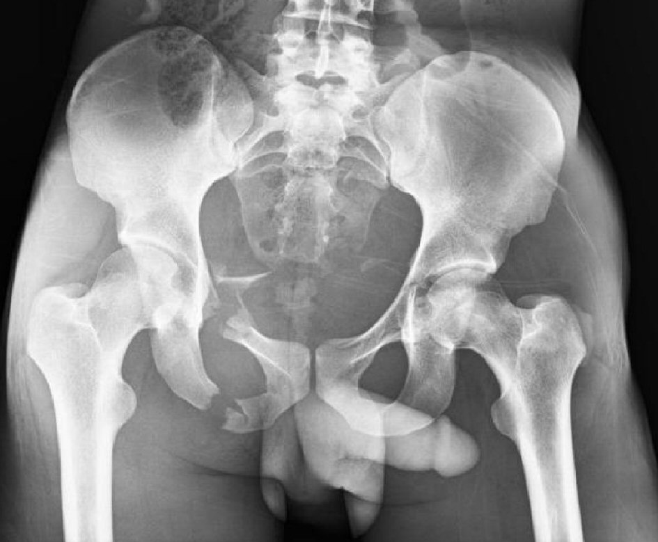
Figure 1. Pelvic X-ray showing fractures of the right superior and inferior rami, left superior ramus, and left acetabulum.
| Journal of Current Surgery, ISSN 1927-1298 print, 1927-1301 online, Open Access |
| Article copyright, the authors; Journal compilation copyright, J Curr Surg and Elmer Press Inc |
| Journal website http://www.currentsurgery.org |
Case Report
Volume 8, Number 1-2, June 2018, pages 13-17
Intra-Vascular Occlusion of the Aorta for Massive Pelvic Trauma: A New Application
Figures