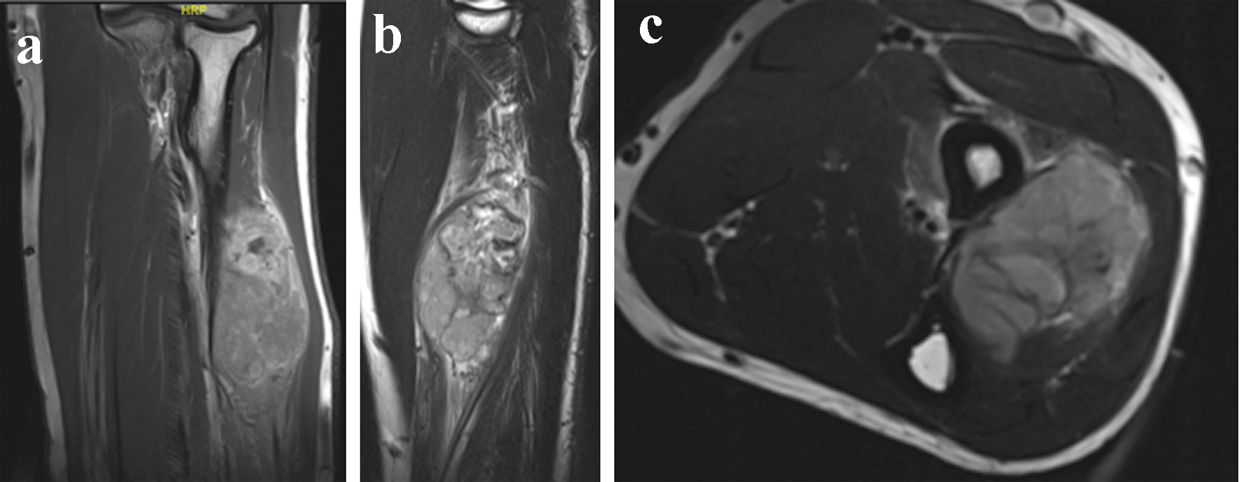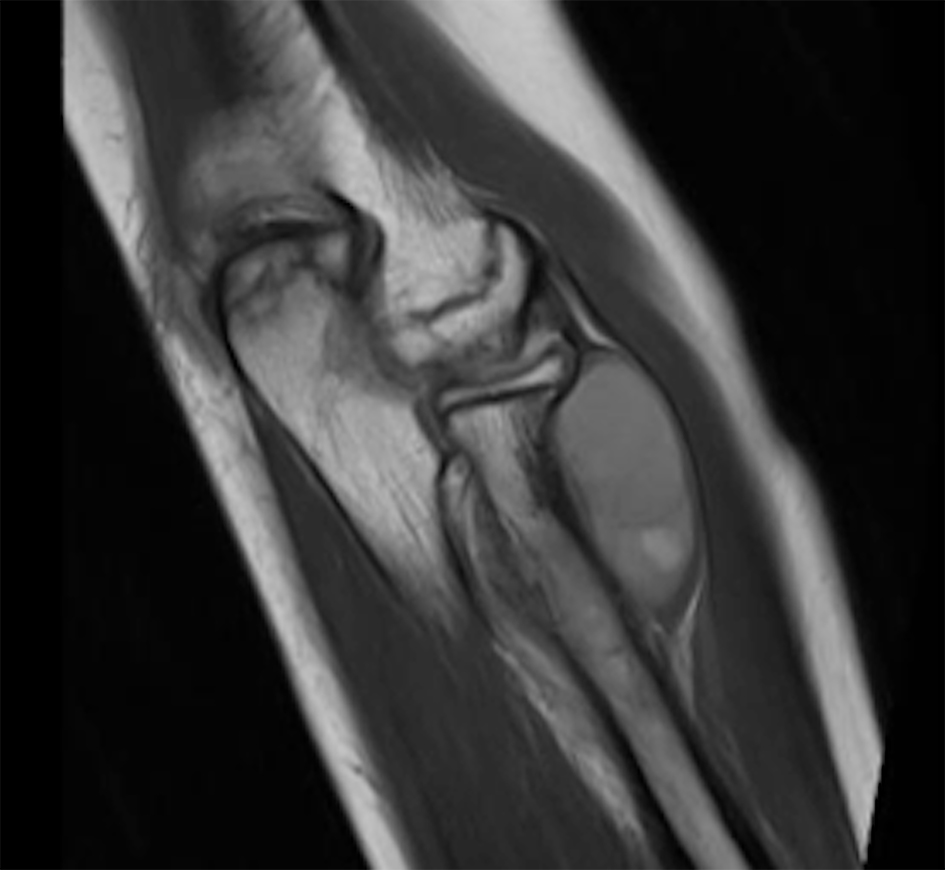
Figure 1. Magnetic resonance imaging of the forearm with a mass measuring 3.2 × 3 × 4.1 cm (coronal cut).
| Journal of Current Surgery, ISSN 1927-1298 print, 1927-1301 online, Open Access |
| Article copyright, the authors; Journal compilation copyright, J Curr Surg and Elmer Press Inc |
| Journal website http://www.currentsurgery.org |
Case Report
Volume 9, Number 2-3, September 2019, pages 32-37
Proximal Radius Ewing’s Sarcoma Resection Followed by Migration of the Proximal Radius: Report of Two Cases
Figures