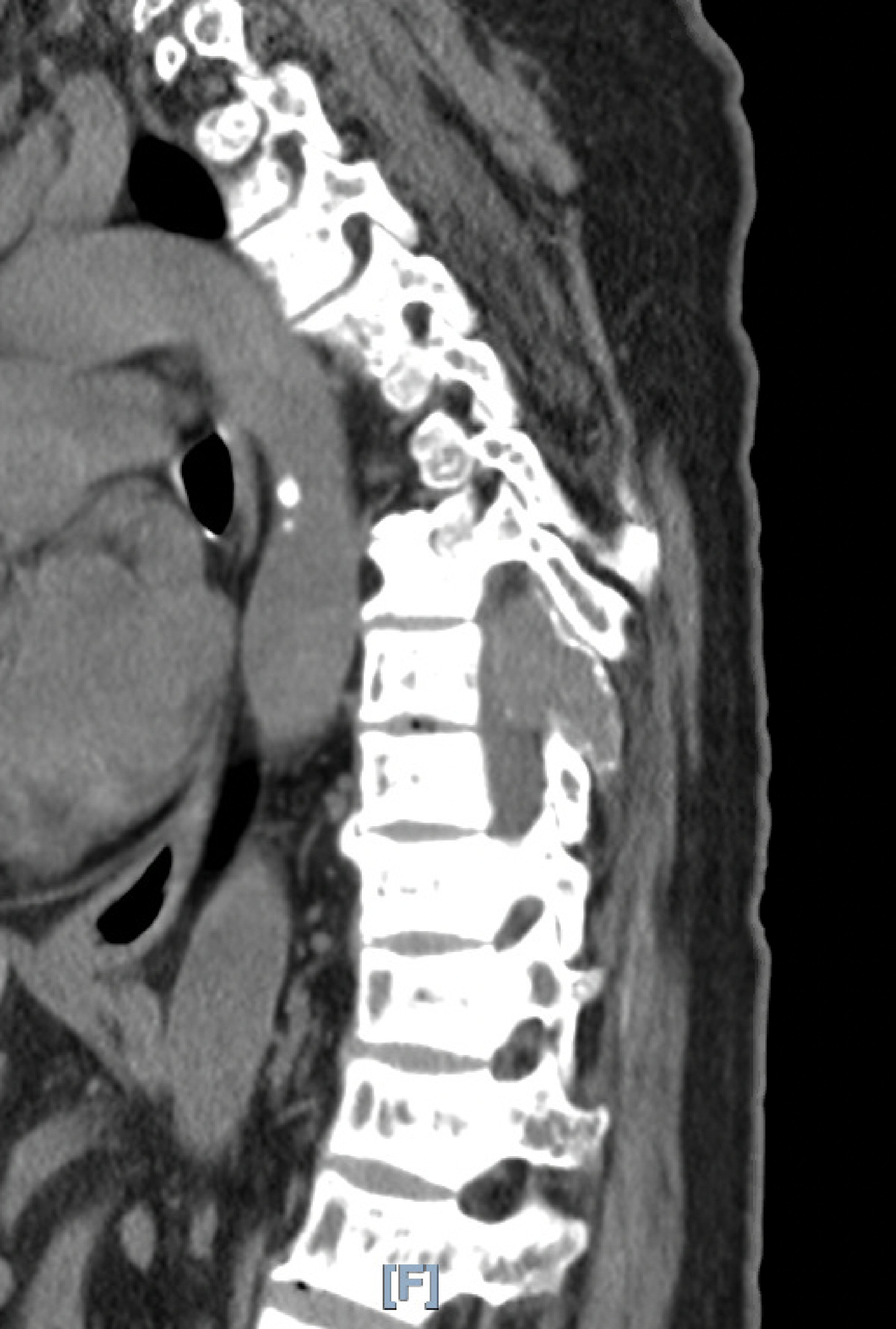
Figure 1. Characteristic CT image of the tumor. CT: computed tomography.
| Journal of Current Surgery, ISSN 1927-1298 print, 1927-1301 online, Open Access |
| Article copyright, the authors; Journal compilation copyright, J Curr Surg and Elmer Press Inc |
| Journal website http://www.currentsurgery.org |
Case Report
Volume 10, Number 1-2, April 2020, pages 13-16
A Rare Presentation of a Brown Tumor on the Spinal Column Secondary to Chronic Renal Failure Managed by Dialysis
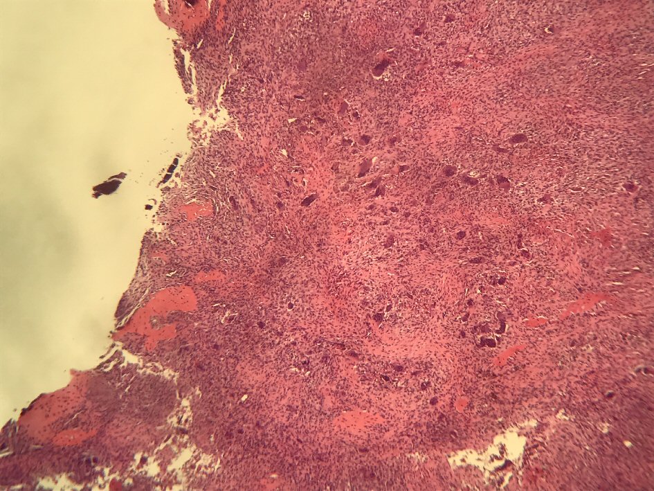
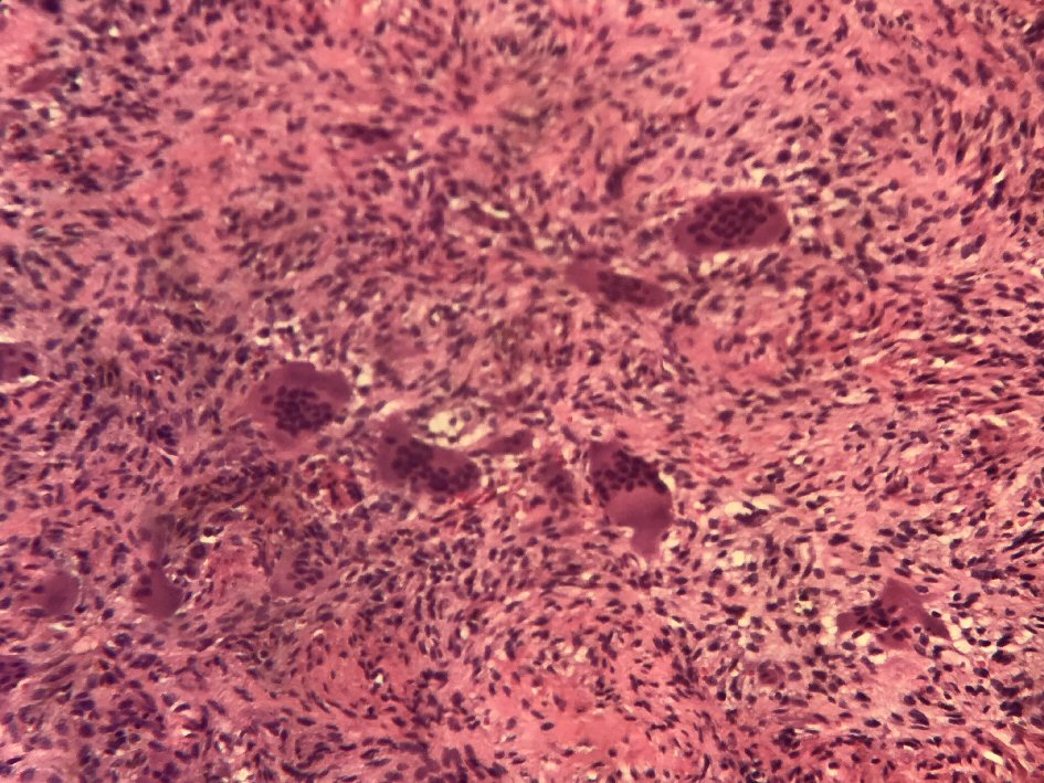
Figures