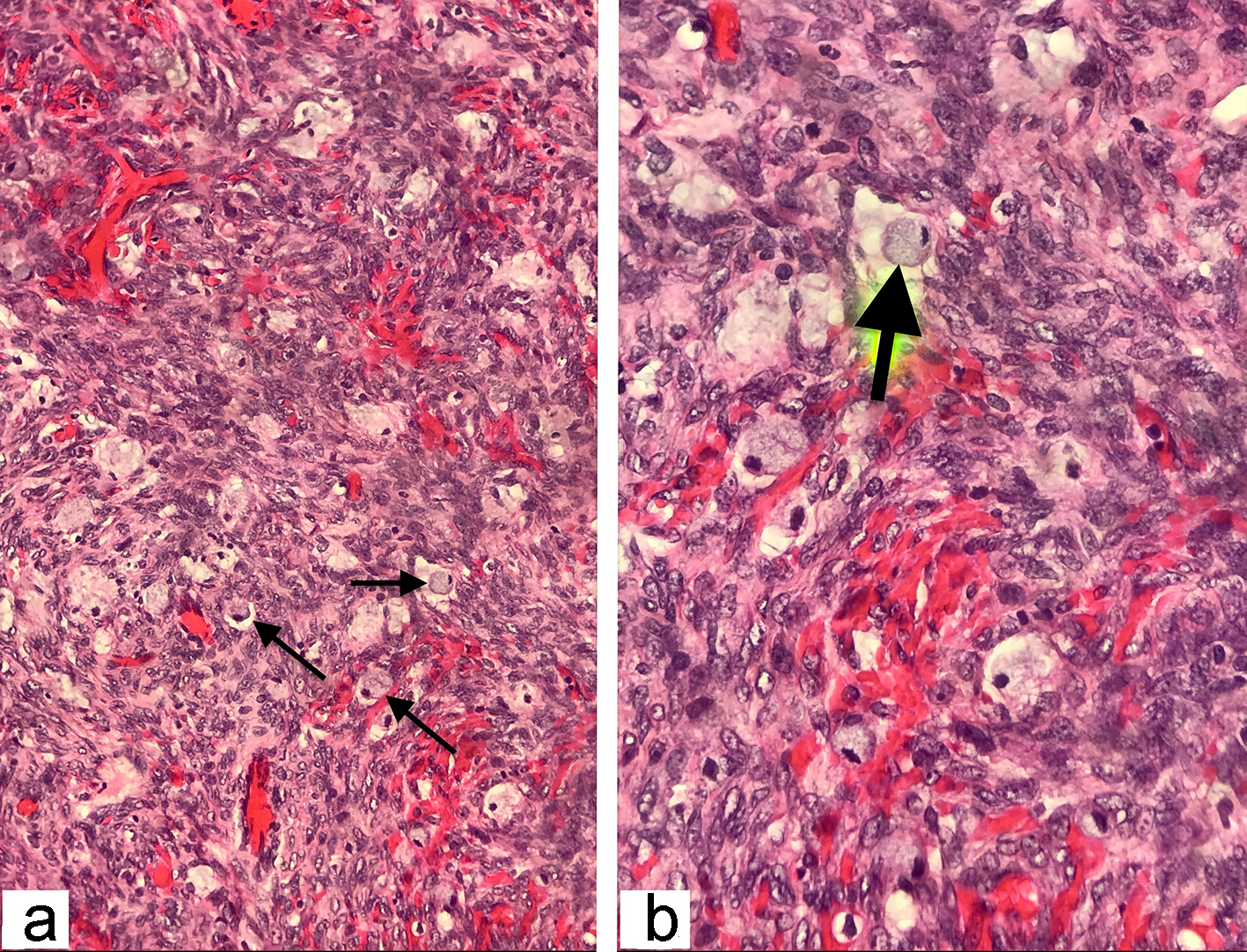
Figure 2. The histopathological examination of the frozen section ovarian-biopsy showing poorly differentiated, diffusely infiltrating signet ring cell carcinoma. Arrows pointing towards the signet ring cells, containing large mucin filled intracytoplasmic vacuoles, with eccentric hyperchromatic nuclei pushed towards the periphery (hematoxylin and eosin staining; magnification: (a) × 200 and (b) × 400).
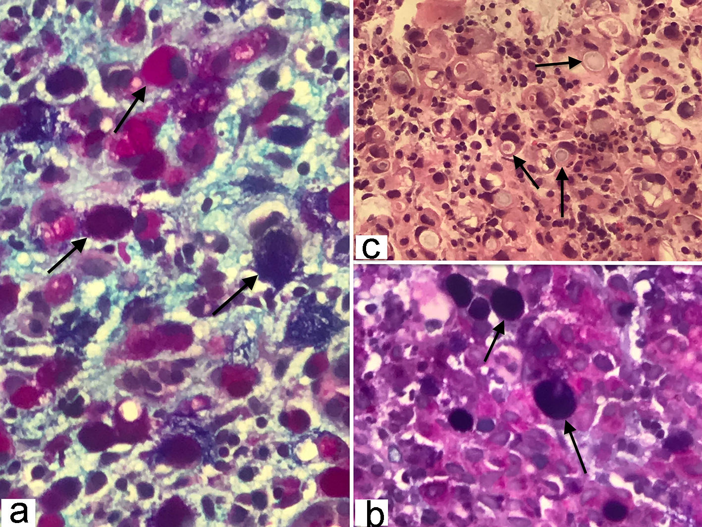
Figure 3. (a, b) Gastric signet-ring cell carcinoma characterized by the presence of abundant intracytoplasmic mucin, combination of Alcian blue (pH 2.5) and periodic acid-Schiff (PAS) stain color the neutral and acid mucins demonstrating a dark blue/purple or magenta coloration (original magnification, × 400). (c) Histopathology of gastric adenocarcinoma showing mucin filled vacuoles that push the nucleus to one side, as shown by the arrows, characteristic of signet ring cell pattern (hematoxylin and eosin, × 200).
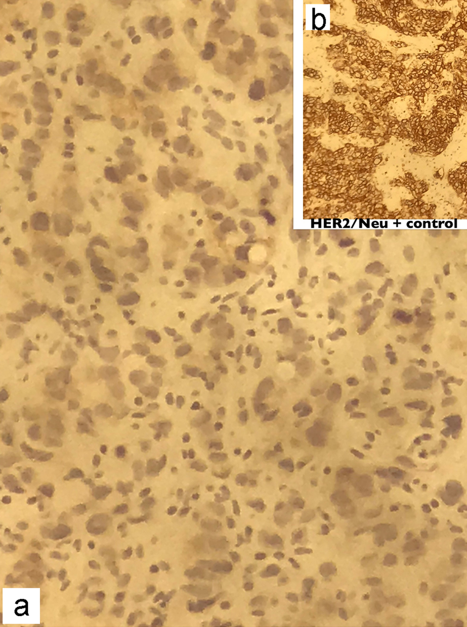
Figure 4. Immunohistochemical staining of the gastric biopsy with Her-2/Neu monoclonal antibodies showing absence of membranous staining in the patient (a), compared to the strong continuous membranous staining in positive control (b). Her-2: human epidermal growth factor receptor 2.