Journal of Current Surgery, ISSN 1927-1298 print, 1927-1301 online, Open Access
Article copyright, the authors; Journal compilation copyright, J Curr Surg and Elmer Press Inc
Journal website https://www.currentsurgery.org

Case Report

Volume 10, Number 3, September 2020, pages 49-53


A Case of Pseudomyxoma Peritonei With a Synchronous Inguinal Hernia

Figures

Figure 1.
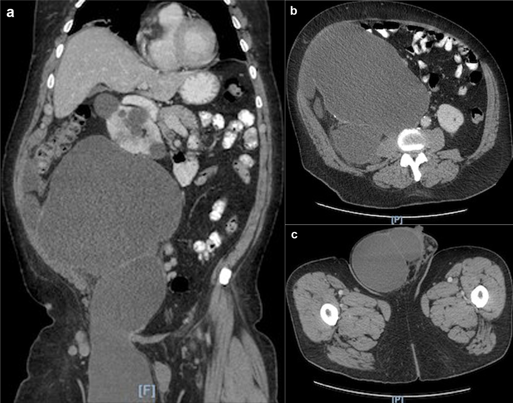
Figure 1. (a) CT scan showing large right retroperitoneal mass extending into the right scrotum. (b) CT scan axial view showing large right retroperitoneal mass. (c) CT scan axial view showing the extension of the retroperitoneal mass into the right scrotum. CT: computed tomography.
Figure 2.
Figure 2. (a) Intraoperative photo of the mucinous ascites seen upon initial entry into peritoneal cavity. (b) Intraoperative photo of resected distal ileum densely adhered to cystic material.
Figure 3.
Figure 3. Umbilical nodule showing dissecting mucin.
Figure 4.
Figure 4. (a) Low-power view of mass showing abundant mucin and a few fragments of neoplastic mucinous glandular epithelium floating within the mucin. (b) High-power view of mass showing abundant mucin and a few fragments of neoplastic mucinous glandular epithelium floating within the mucin.