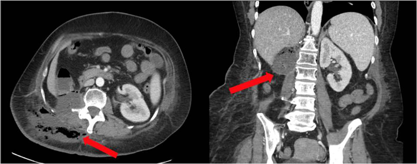
Figure 1. CT of the abdomen and pelvis with contrast showing a complex fluid collection with air in the right renal fossa (red arrows) extending into the subcutaneous tissues of the back. CT: computed tomography.
| Journal of Current Surgery, ISSN 1927-1298 print, 1927-1301 online, Open Access |
| Article copyright, the authors; Journal compilation copyright, J Curr Surg and Elmer Press Inc |
| Journal website https://www.currentsurgery.org |
Case Report
Volume 10, Number 3, September 2020, pages 54-58
Colonic Perforation Associated with Necrotizing Fasciitis in a Patient Receiving Tyrosine Kinase Inhibitor (Pazopanib) for Recurrent Retroperitoneal Renal Cell Carcinoma
Figures