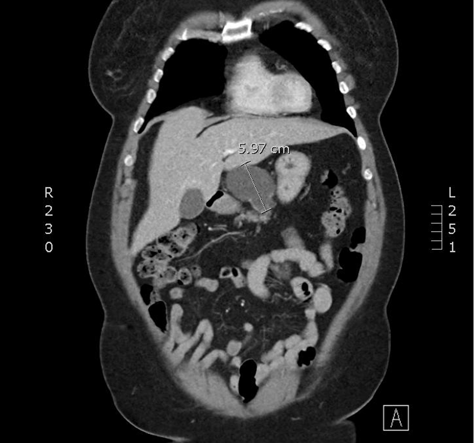
Figure 1. An axial slice from a CT scan with contrast. A cystic mass with few thin septae suggesting multi-loculation associated with the superior and anterior pancreatic body. CT: computed tomography.
| Journal of Current Surgery, ISSN 1927-1298 print, 1927-1301 online, Open Access |
| Article copyright, the authors; Journal compilation copyright, J Curr Surg and Elmer Press Inc |
| Journal website https://www.currentsurgery.org |
Case Report
Volume 11, Number 1, March 2021, pages 24-27
Pancreatic Cystic Lymphangioma: Case Report and Literature Review
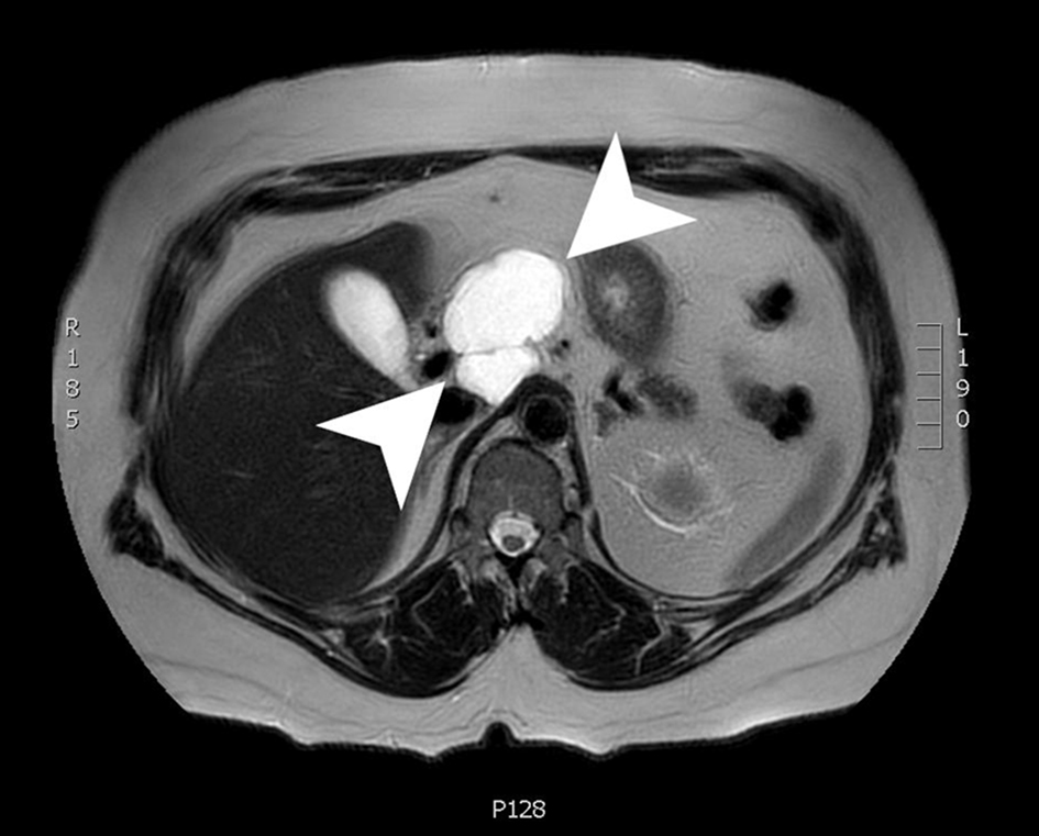
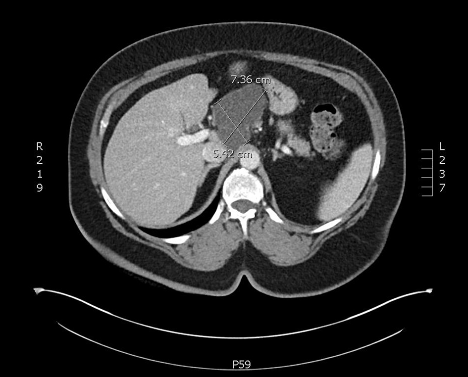
Figures