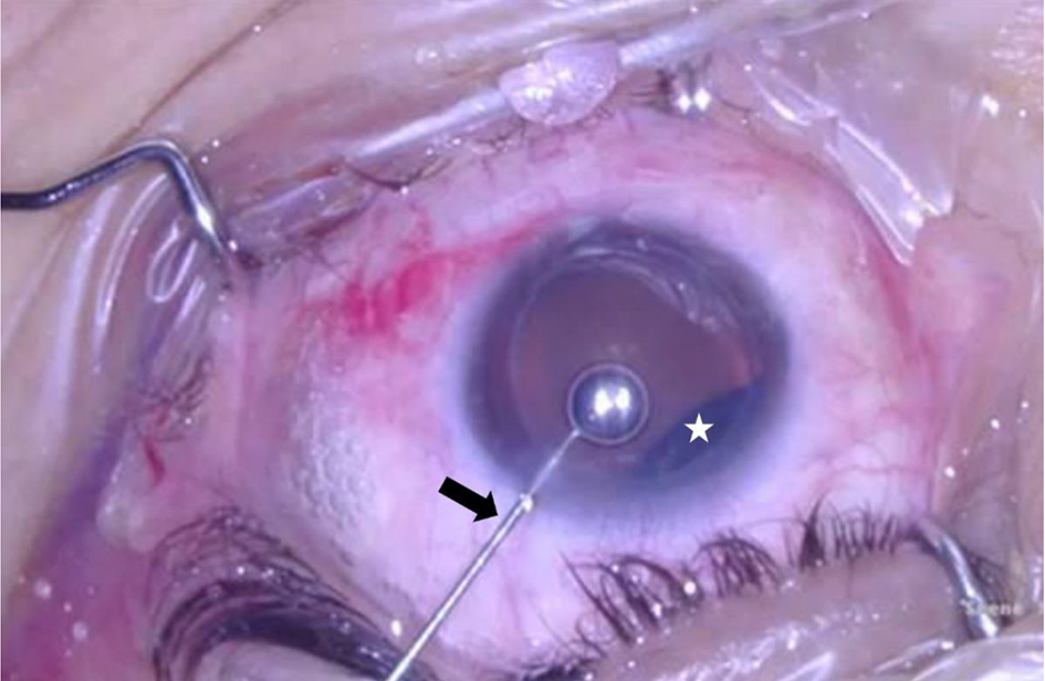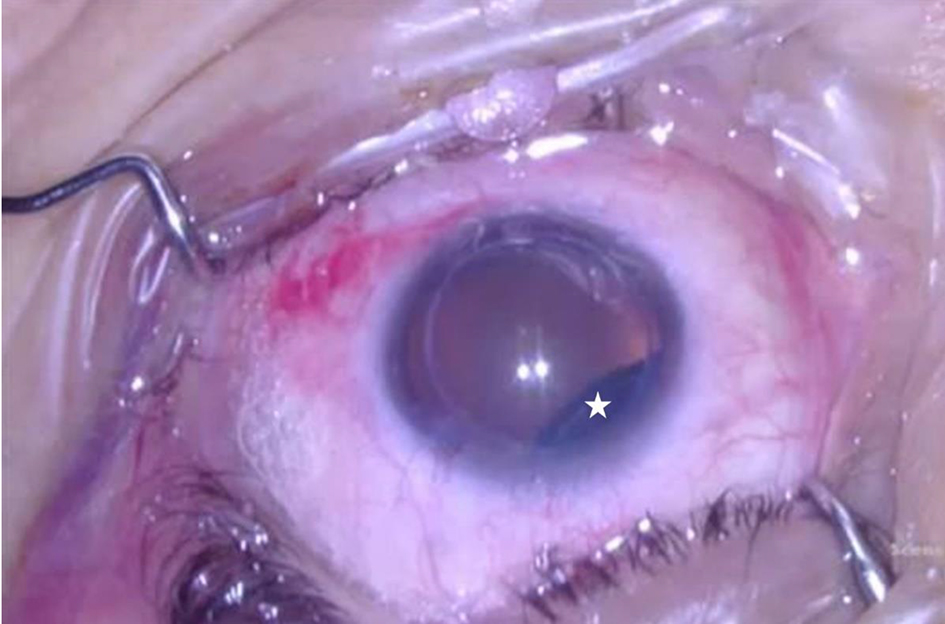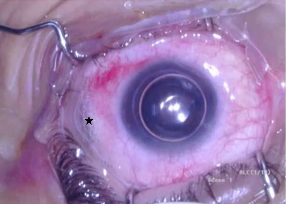
Figure 1. Gas filling the anterior chamber after the drainage of aqueous. Surgeon’s view, left eye. Note a small amount of subconjunctival gas in the site of injection (indicated by the black star).
| Journal of Current Surgery, ISSN 1927-1298 print, 1927-1301 online, Open Access |
| Article copyright, the authors; Journal compilation copyright, J Curr Surg and Elmer Press Inc |
| Journal website https://www.currentsurgery.org |
Case Report
Volume 11, Number 1, March 2021, pages 21-23
A Rare Case of Anterior Chamber SF6 Gas as a Complication of Pneumatic Retinopexy
Figures