Journal of Current Surgery, ISSN 1927-1298 print, 1927-1301 online, Open Access
Article copyright, the authors; Journal compilation copyright, J Curr Surg and Elmer Press Inc
Journal website https://www.currentsurgery.org

Case Report

Volume 11, Number 3, September 2021, pages 65-68


Laparoscopic Treatment of Liver Abscess by a Fishbone Impaction: A Case Report

Figures

Figure 1.
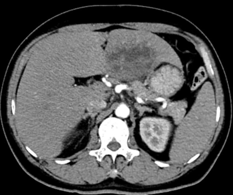
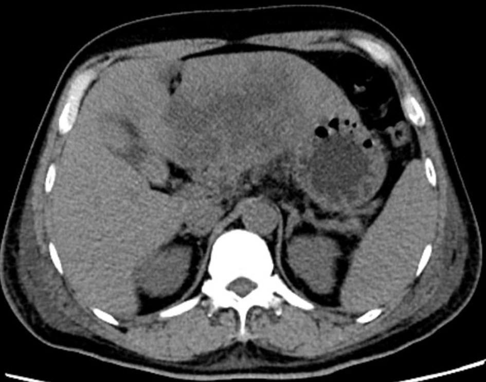
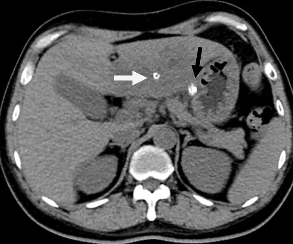
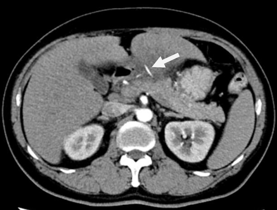
Figure 1. Enhanced computed tomography (CT) scan of the upper abdomen demonstrating the needle-shaped dense shadow (arrow) between the stomach and the liver.
Figure 2.
Figure 2. Enhanced computed tomography (CT) scan of the upper abdomen showing a 7 cm hypodense mass in the left hepatic lobe.
Figure 3.
Figure 3. Plan computed tomography (CT) scan of the upper abdomen showing liquefaction within liver abscess after antibody therapy for 4 days before operation.
Figure 4.
Figure 4. The hepatic abscess in the left hepatic lobe was opened by ultrasonic scalpel.
Figure 5.
Figure 5. Removal of the fishbone in the omentulum around the hepatoduodenal ligament by forceps.
Figure 6.
Figure 6. The foreign body removed: a 2.5 cm long fishbone.
Figure 7.
Figure 7. Plan computed tomography (CT) scan of the upper abdomen after operation showing the drainage pipe in the abscess cavity of liver (white arrow) and the drainage pipe out of the abscess cavity of the liver (black arrow).