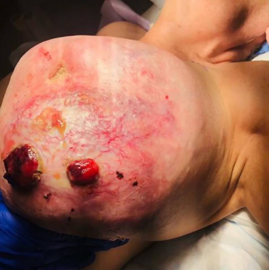
Figure 1. Pre-operative phyllodes tumor of the left breast.
| Journal of Current Surgery, ISSN 1927-1298 print, 1927-1301 online, Open Access |
| Article copyright, the authors; Journal compilation copyright, J Curr Surg and Elmer Press Inc |
| Journal website https://www.currentsurgery.org |
Case Report
Volume 11, Number 3, September 2021, pages 69-72
Giant Borderline Phyllodes Tumor With Malignant Presentation: A Case Report
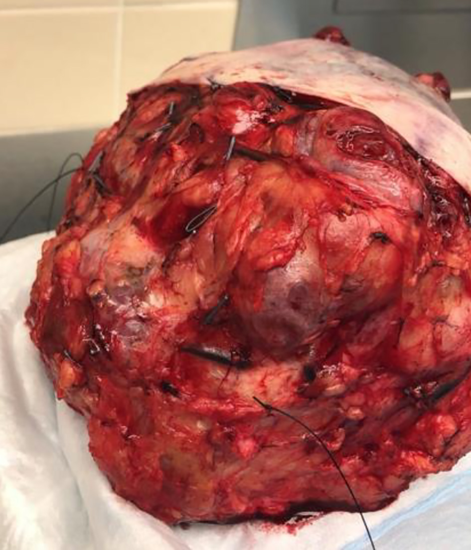
Figures
Table
| Histological features | Benign | Borderline | Malignant |
|---|---|---|---|
| HPF: high-power field. | |||
| Stromal hypercellularity | Minimal | Moderate | Marked |
| Cellular pleomorphism | Little | Moderate | Marked |
| Mitosis | 0 - 4/10 HPF | 5 - 9/10 HPF | > 10/10 HPF |
| Margins | Pushing | Zone of microscopic invasion around tumor margins | Invasive |
| Stromal pattern | Uniform stromal distribution | Heterogenous stromal expansion | Marked stromal overgrowth |
| Heterologous stromal differentiation | Rare | Rare | Not uncommon |