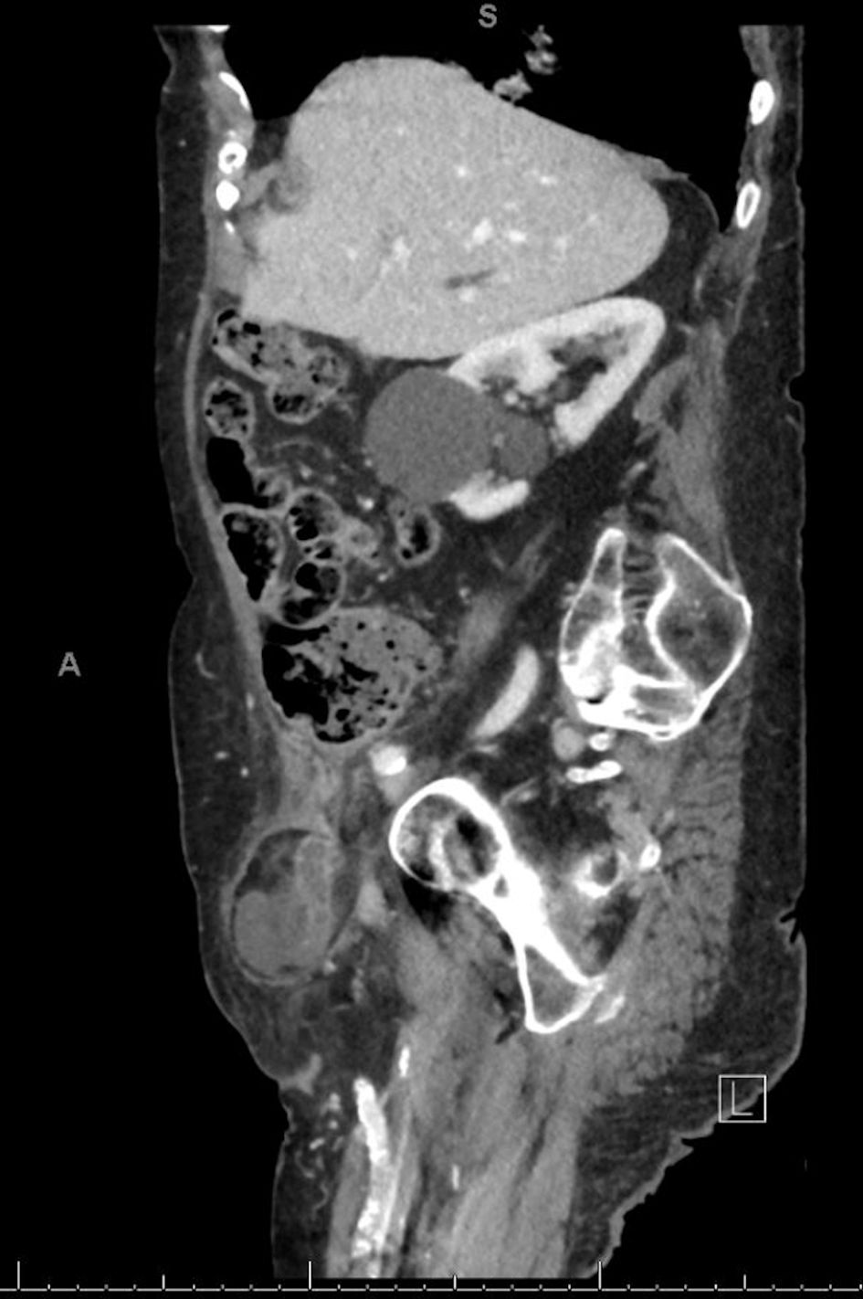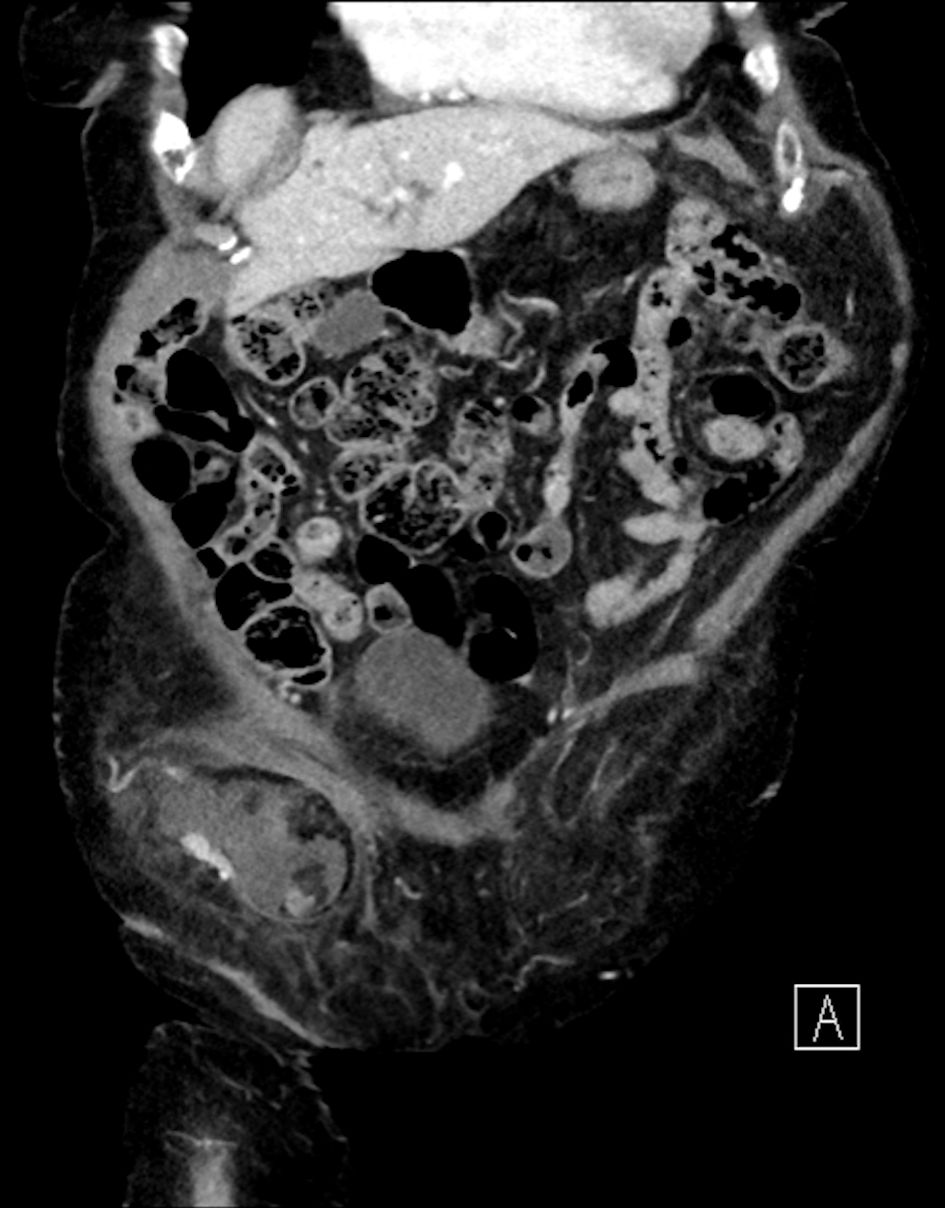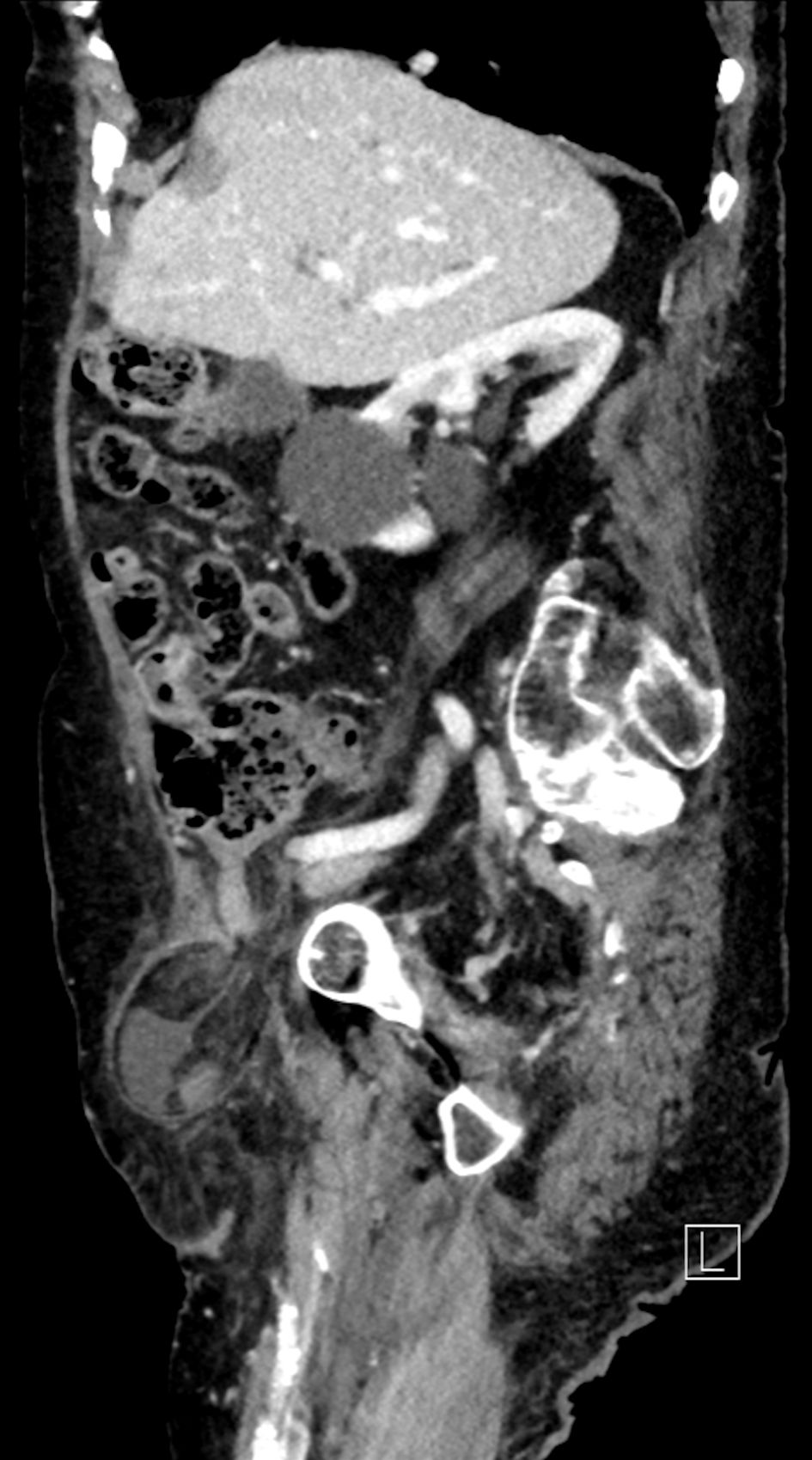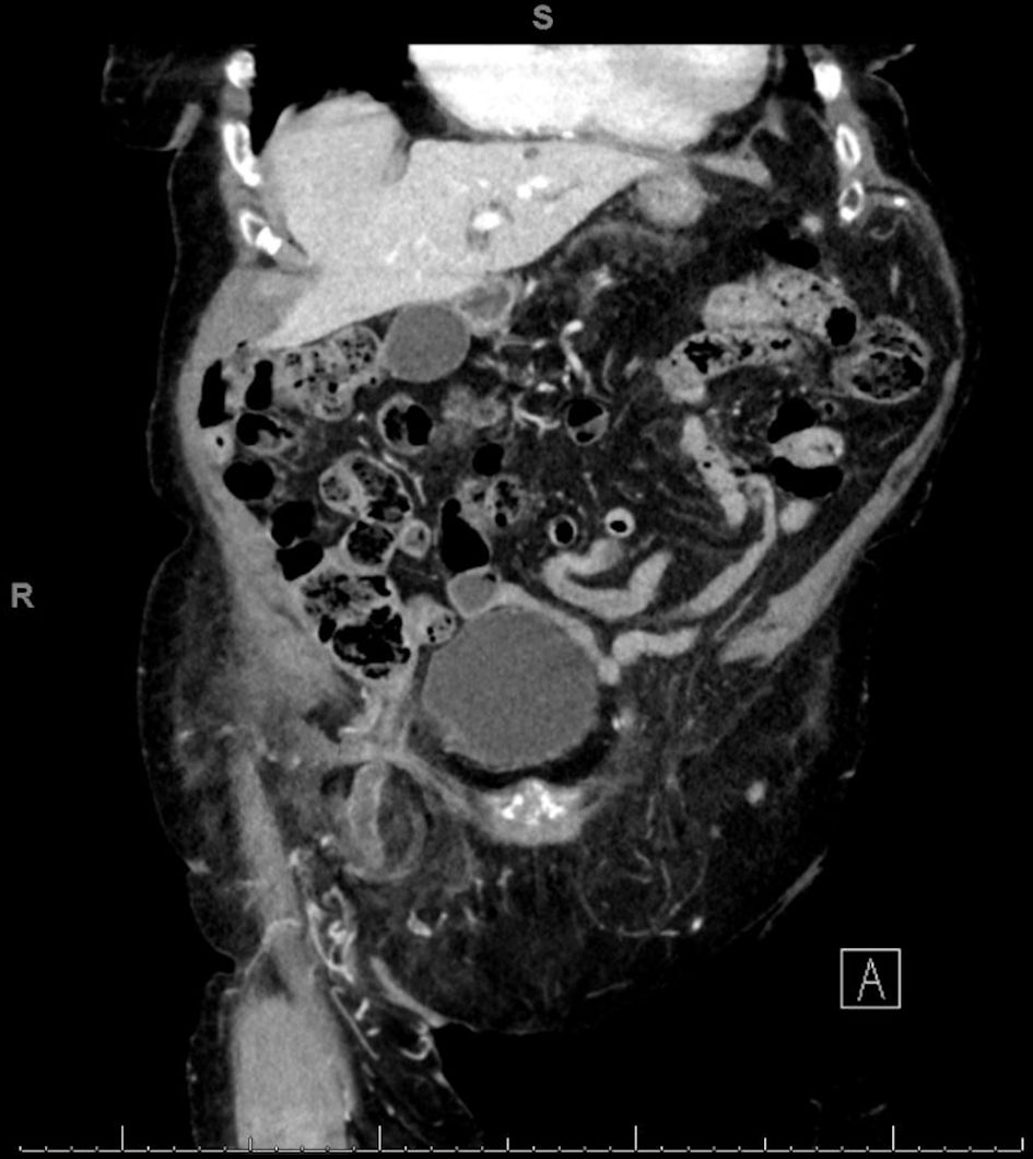
Figure 1. CT coronal scan: right femoral hernia containing liquid effusion and an inflamed cecal appendix. CT: computed tomography.
| Journal of Current Surgery, ISSN 1927-1298 print, 1927-1301 online, Open Access |
| Article copyright, the authors; Journal compilation copyright, J Curr Surg and Elmer Press Inc |
| Journal website https://www.currentsurgery.org |
Case Report
Volume 11, Number 4, December 2021, pages 97-100
Complex de Garengeot’s Hernia With a Bladder Diverticulum
Figures