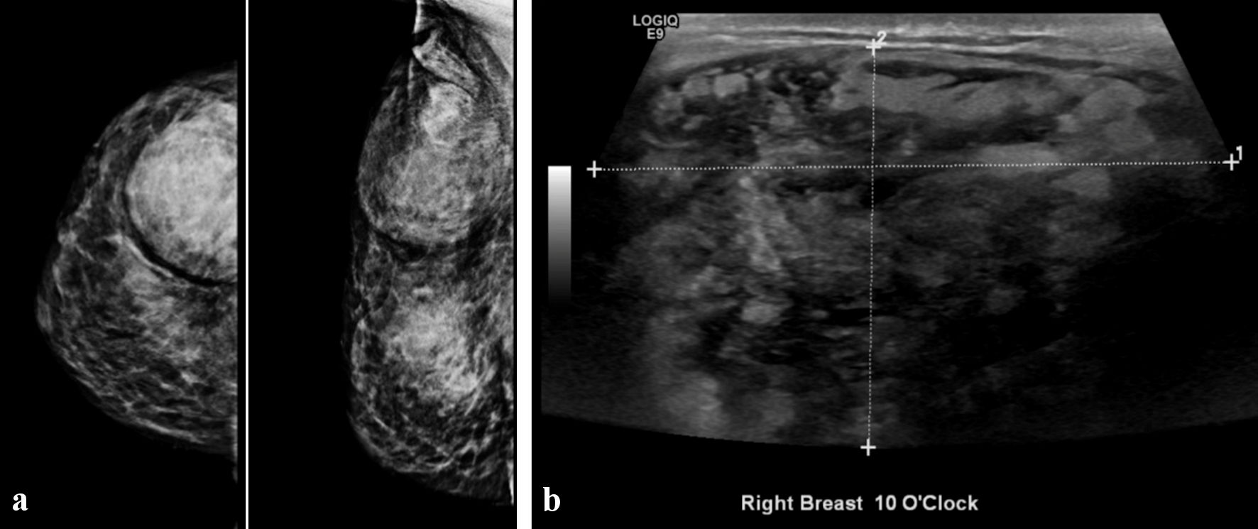
Figure 1. Breast imaging showing circumscribed oval heterogeneous high density mainly hyperechoic mass in the right breast at 10 o’clock. (a) Mammography. (b) Ultrasonography.
| Journal of Current Surgery, ISSN 1927-1298 print, 1927-1301 online, Open Access |
| Article copyright, the authors; Journal compilation copyright, J Curr Surg and Elmer Press Inc |
| Journal website https://www.currentsurgery.org |
Case Report
Volume 12, Number 2, December 2022, pages 45-49
A Large Myofibroblastoma of the Breast in a Premenopausal Woman: A Case Report and Review of the Literature
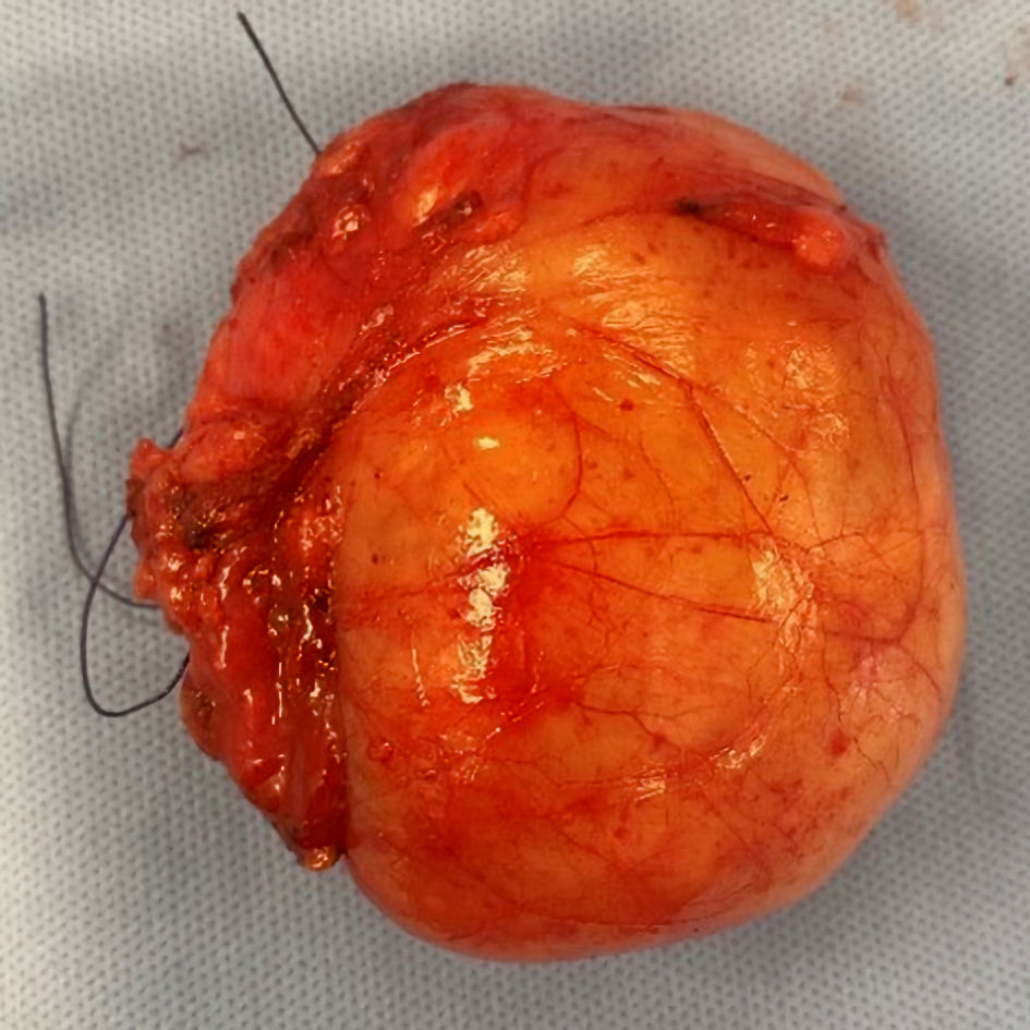
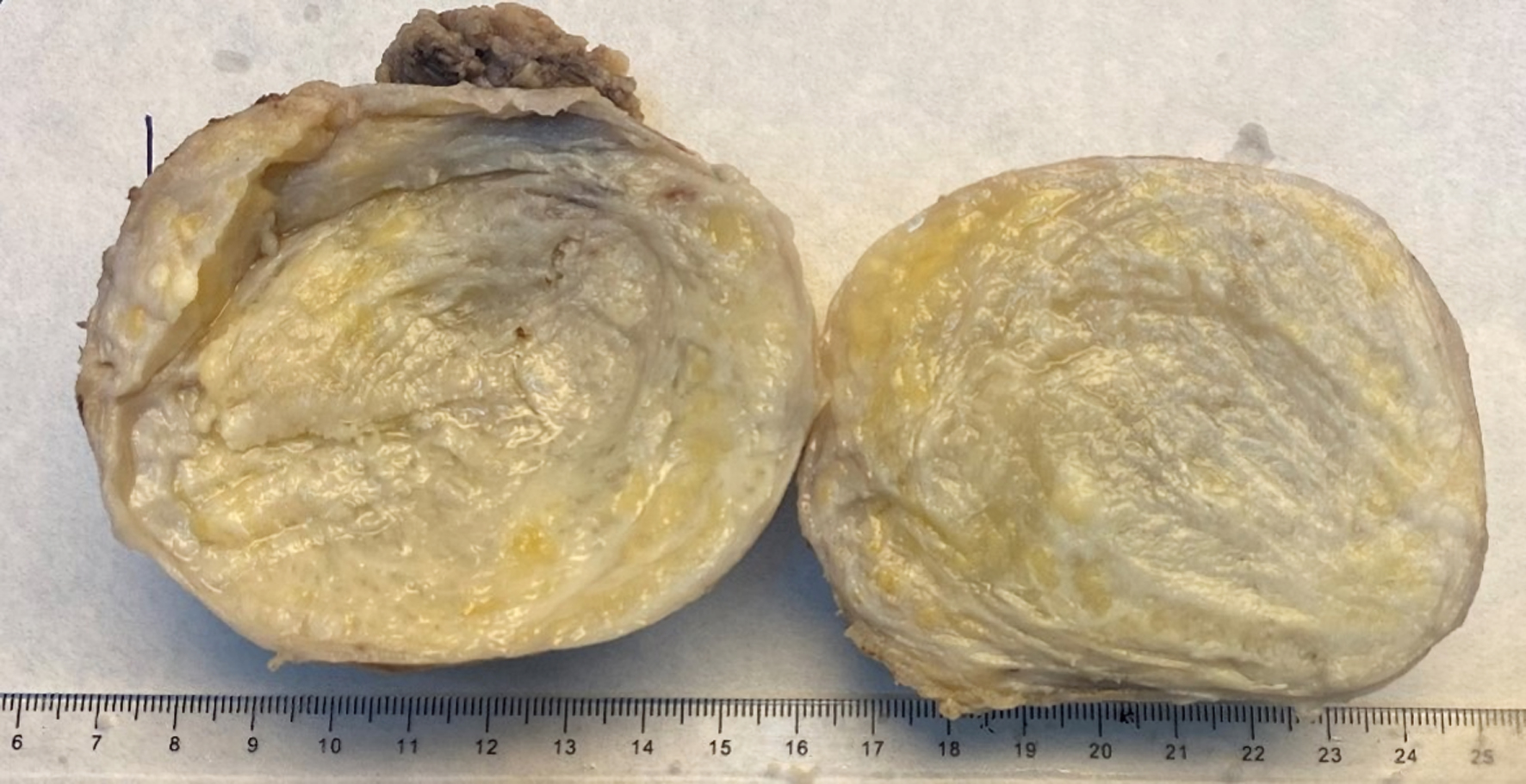
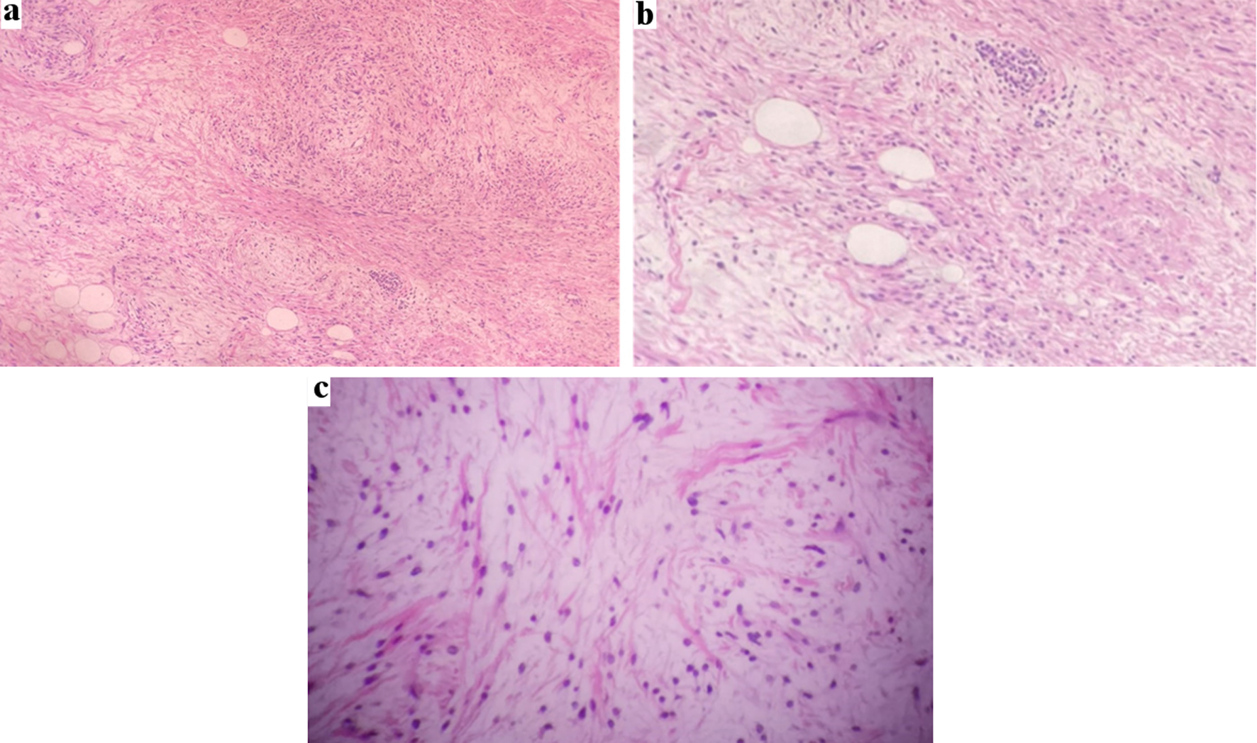
Figures
Table
| Variant | Description |
|---|---|
| MFB: myofibroblastoma. | |
| Cellular | Dense proliferation of spindle cells, usually with infiltrative boarder. |
| Epithelioid | MFB composed exclusively or predominantly (> 50%) of epithelioid cells. |
| Deciduoid-like | Larger cells with copious eosinophilic cytoplasm, vesicular nuclei, with single or multiple prominent nucleoli. |
| Collagenized/fibrous | Highly collagenous stroma. |
| Infiltrative | Invasive growth pattern, entrapping surrounding glandular tissue and fat. |
| Lipomatous | MFB composed predominantly (i.e., > 75% of the entire neoplasm) of adipocytes. |
| Myxoid | Entirely or predominantly myxoid stroma in which spindle cells and stellate cells are embedded. |
| Mixed | Two or more variants coexisting in the same tumor. |