Journal of Current Surgery, ISSN 1927-1298 print, 1927-1301 online, Open Access
Article copyright, the authors; Journal compilation copyright, J Curr Surg and Elmer Press Inc
Journal website https://www.currentsurgery.org

Short Communication

Volume 13, Number 1, September 2023, pages 6-11


The Effects of Cisplatin on Gastrostomy Site Healing

Figures

Figure 1.
Figure 1. Gastrostomy tube insertion. The figure represents the technique that the gastrostomy placement was performed. Left flank incision was made (a) to place the tube through (b). The tube was placed into stomach and fixed to the lateral abdominal wall (c).
Figure 2.
Figure 2. Masson’s trichrome staining showed more collagen formation in surgical site tissue at 17 versus 7 days post-cisplatin. The arrows heads show the collagen fibers.
Figure 3.
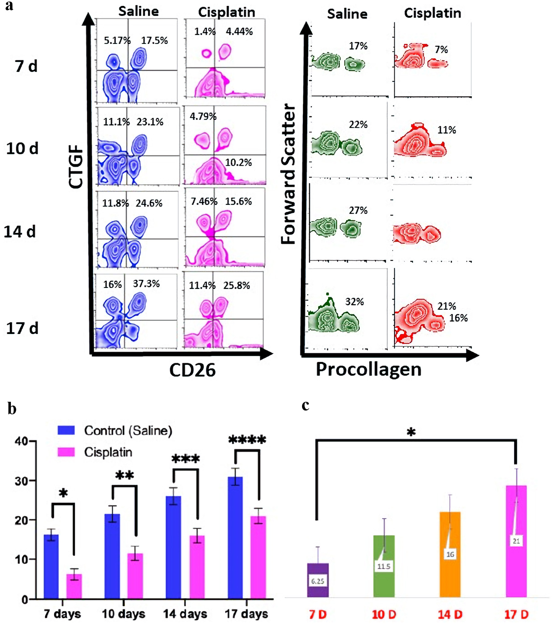
Figure 3. The flow cytometry for the CTGF, CD26, and procollagen expression from day 7 to 17. Levels at 14 days post-cisplatin were comparable to levels at 7 days post-saline (a). The statistical analyses were performed using two-way ANOVA with Tukey multiple comparison test. *P < 0.05; **P < 0.01; ***P < 0.001; ****P < 0.0001. The graph shows the levels of procollagen type 1 by flow cytometry were a significant higher expression that was present in the control group compared to the treatment group (P < 0.0001) for each time point (b). There was a statistically difference in procollagen type 1 between the 7 days post-chemotherapy group and the 17 days post-chemotherapy group for each time point (c). CTGF: connective tissue growth factor; ANOVA: analysis of variance.