Figures
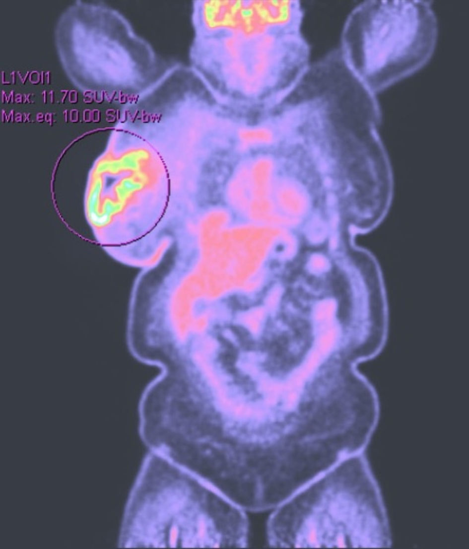
Figure 1. PET scan at 4 months revealing increased tracer uptake in the right breast. PET: positron emission tomography.
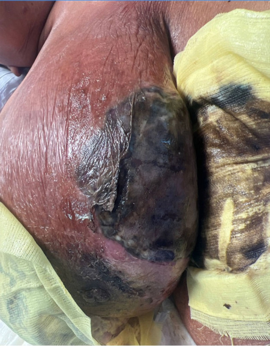
Figure 2. The image illustrating the condition of the breast upon arrival at the emergency room, characterized by intense erythema, skin de-epithelialization, ischemic changes, and tissue necrosis.
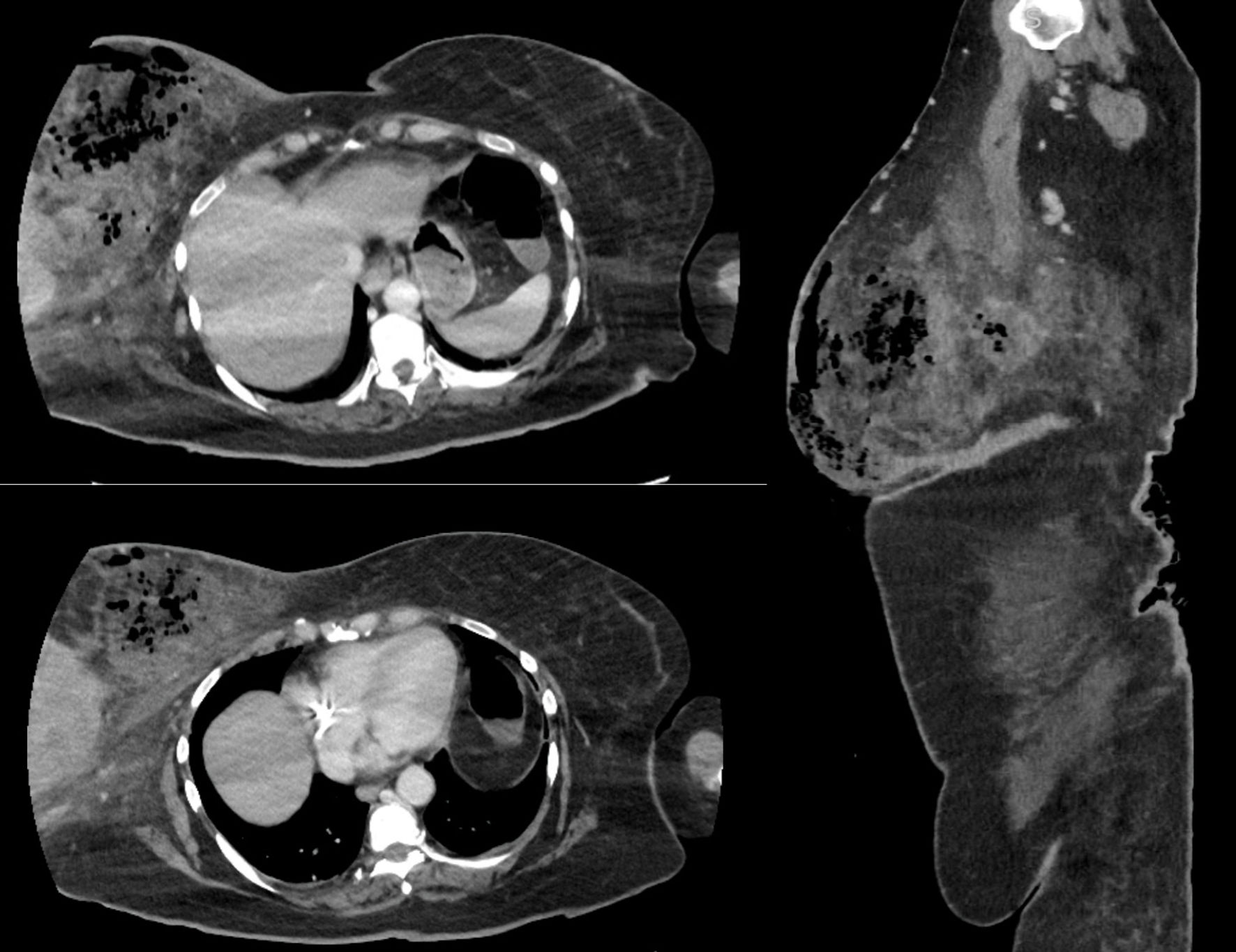
Figure 3. CT scan of the chest and abdominopelvic of the breast necrotizing infection. This image illustrates large pockets of gas within the right breast parenchyma with edema. CT: computed tomography.
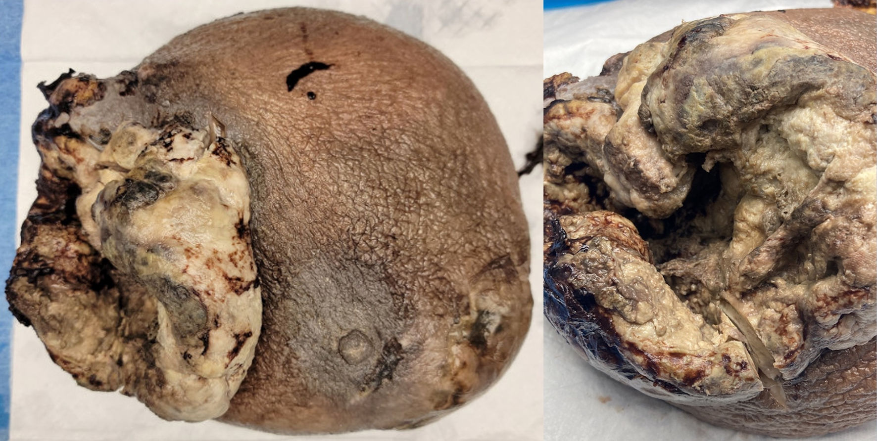
Figure 4. Ulcerated and necrotic mass with cavitation involving breast tissue (gross pathology picture).
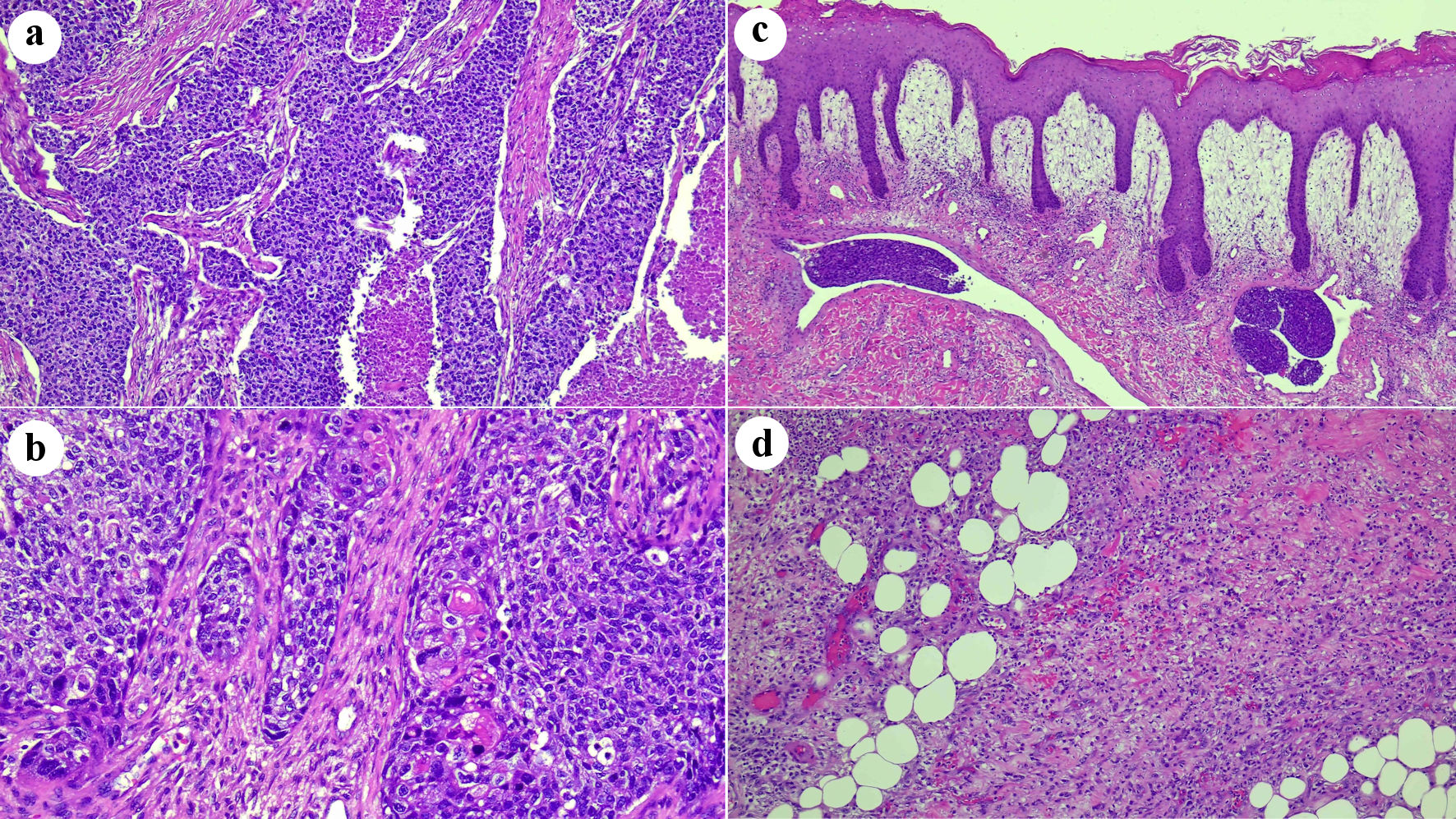
Figure 5. (a) Histologic features of invasive poorly differentiated ductal carcinoma. Sheets of atypical cells with areas of necrosis (original magnification × 100). (b) Scattered areas with squamous morules, compatible with an invasive ductal carcinoma with metaplastic squamous cell features (original magnification × 200). (c) Overlying skin with abundant dermal edema, acute inflammation, and tumor emboli in dermal lymphatic vessels (original magnification × 50). (d) Underlying fatty breast tissue with necrosis and abundant acute inflammation (original magnification × 10).