Journal of Current Surgery, ISSN 1927-1298 print, 1927-1301 online, Open Access
Article copyright, the authors; Journal compilation copyright, J Curr Surg and Elmer Press Inc
Journal website http://www.currentsurgery.org

Case Report

Volume 2, Number 3, June 2012, pages 113-115


Breast MRI as a Problem Solving Tool in Paget’s Disease of the Nipple

Figures

Figure 1.
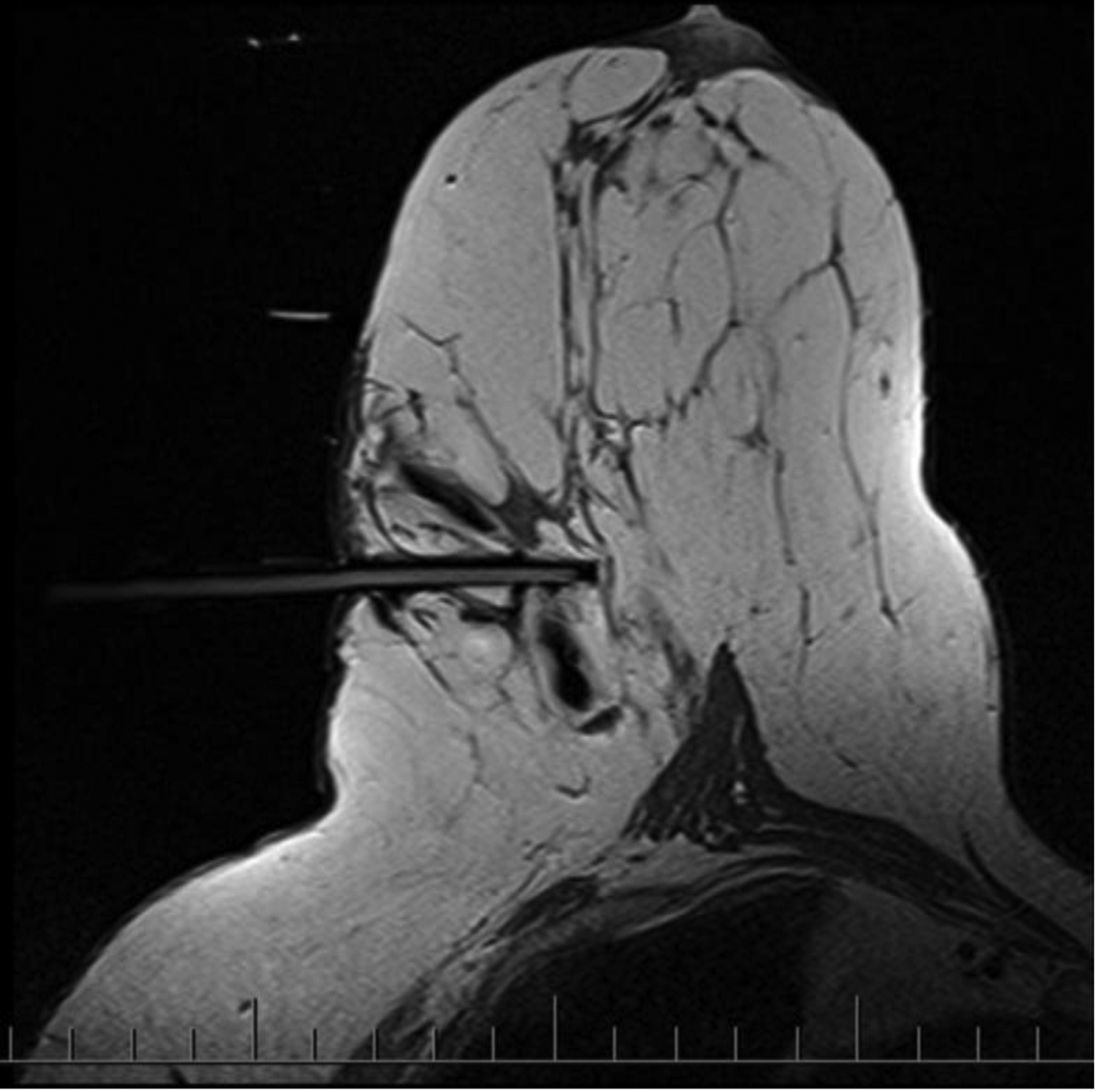
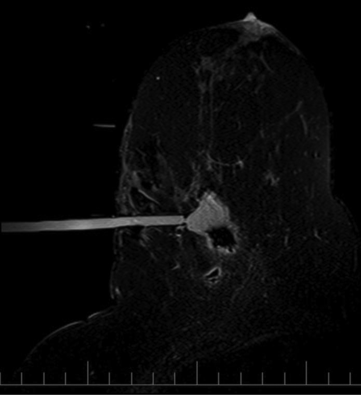
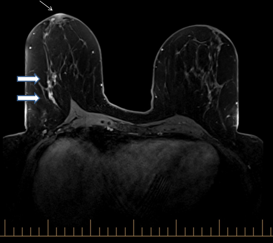
Figure 1. Axial fat-supressed T1-weighted image showing non-mass-like enhancement in the outer central right breast, prepectoral region (large arrows) and unilateral nipple enhancement on the right, in keeping with the known diagnosis of Paget’s disease of the nipple (small arrow).
Figure 2.
Figure 2. Axial non-fat-supressed T1-weighted image with MRI visible obturator in position, confirming correct needle position.
Figure 3.
Figure 3. Axial fat-supressed T1-weighted image post biopsy confirms a cavity in the area of previous non-mass like enhancement in the outer central right breast, prepectoral region.