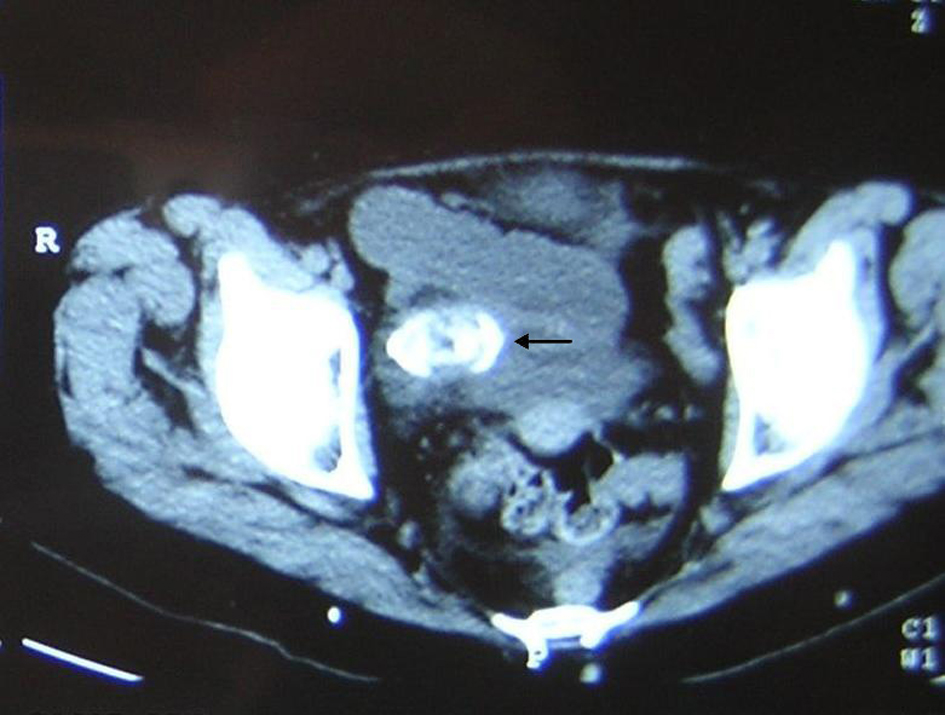
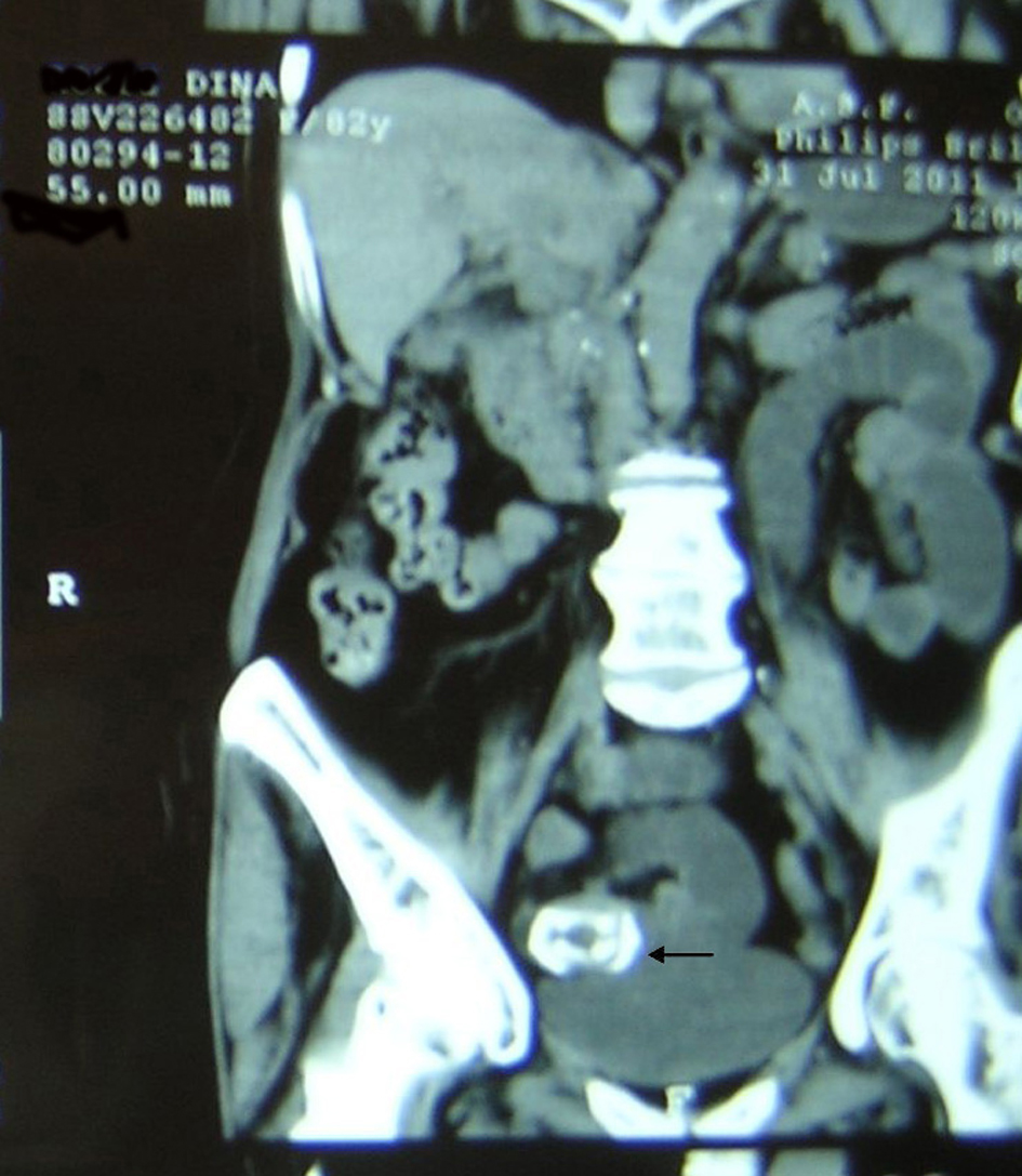
Figure 1. Abdominal tomography shows ectopic stone in the ileum.
| Journal of Current Surgery, ISSN 1927-1298 print, 1927-1301 online, Open Access |
| Article copyright, the authors; Journal compilation copyright, J Curr Surg and Elmer Press Inc |
| Journal website http://www.currentsurgery.org |
Original Article
Volume 2, Number 3, June 2012, pages 84-88
Gallstone Ileus: Diagnostic and Surgical Dilemma
Figures
Table
| Patient | Sex | Age | Concomitant medical conditions | History of cholelithiasys | ASA Score | Radiological findings | Preoperative diagnosis | Site of stone impaction/diame-ter/Surgical treat-ment/duration of surgery | Postoperative Course/hospital stay (days) |
|---|---|---|---|---|---|---|---|---|---|
| S.O. | F | 86 | Diabetes mellitus; Arterial hypertension | - | 4 | Pneumobilia + Air-fluid level + Detection of stone + | Gallstone ileus | Jejunum / 4 cm / enterolithotomy / 30 min | Heart failure / Death on the 14th post-op |
| G.S. | F | 84 | Diabetes mellitus; Arterial hypertension | + | 3 | Pneumobilia + Air-fluid level + Detection of stone - | Intestinal obstruction | Jejunum / ? / Enterolithotomy / 45 min | Uneventful / 9 days |
| F.M. | F | 75 | Atrial fibrillation | + | 3 | Pneumobilia - Air-fluid level + Detection of stone - | Intestinal obstruction | Jejunum / 4 cm / enterolithotomy cholecystectomy fistula closure / 85 min | Uneventful / 8 days |
| R.D. | F | 82 | - | + | 2 | Pneumobilia + Air-fluid level + Detection of stone + | Gallstone ileus | Ileum / 5 cm / enterolithotomy / 30 min | Uneventful / 11 days |