Journal of Current Surgery, ISSN 1927-1298 print, 1927-1301 online, Open Access
Article copyright, the authors; Journal compilation copyright, J Curr Surg and Elmer Press Inc
Journal website http://www.currentsurgery.org

Case Report

Volume 2, Number 4-5, October 2012, pages 149-153


Allergic Fungal Rhinosinusitis in the Lesser and Greater Wings of the Sphenoid With Resultant Optic Neuropathy: A Case Report and Review of the Literature

Figures

Figure 1.
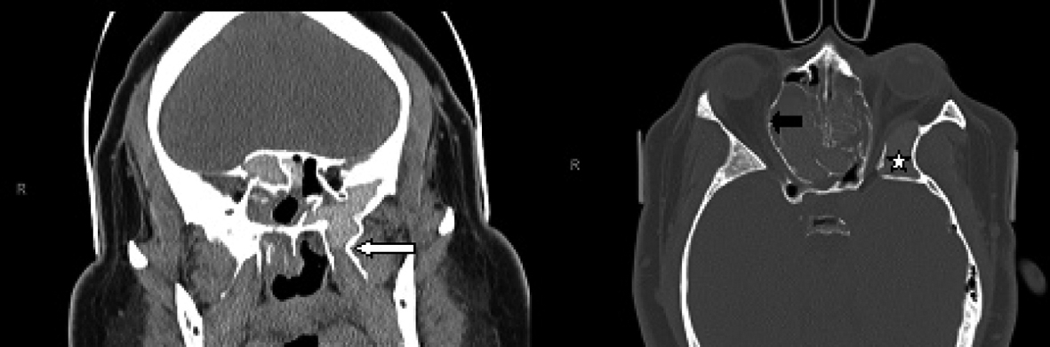
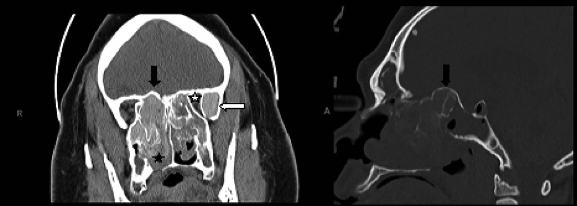
Figure 1. Coronal CT scan of the paranasal sinuses (left image) shows enlarged and opacified left greater wing of sphenoid with high attenuation material (white arrow), along with narrowing of the orbital apex on the left compared to the right side (white star). The fungal hyperattenuation areas also extend intranasally (black star). There is bulging of the roof of the posterior ethmoids into the intracranial space as demonstrated on both the coronal and sagittal CT scans (right image) labeled with black arrows.
Figure 2.
Figure 2. Coronal CT scan of the paranasal sinuses (left image) demonstrates the hyperaerated left sphenoid sinus extending far lateral and inferior with opacification of the left pterygoid (white arrow). The axial CT scan (right image) demonstrates bulging of the lamina papyracea into the orbits, more so on the right side (black arrow). There is redemonstration of the aerated and opacified left greater wing of sphenoid (white star).
Figure 3.
Figure 3. Axial MRI with T1-weighted (left image) and T2-weighted (right image) demonstrating the narrowing of the left orbital apex compared to the right side with impingement of the optic nerve (white arrow). Signal void is demonstrated with the black arrow on the T-2 weighted image.