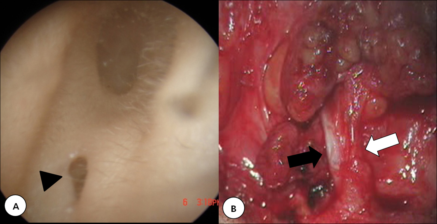
Figure 1. A: A 20-year-old female presented with a sinus opening (arrow head) in the left external auditory canal. B: The track (black arrow) paralleled the normal external auditory canal and terminated deep to the facial nerve (white arrow).
| Journal of Current Surgery, ISSN 1927-1298 print, 1927-1301 online, Open Access |
| Article copyright, the authors; Journal compilation copyright, J Curr Surg and Elmer Press Inc |
| Journal website http://www.currentsurgery.org |
Case Report
Volume 2, Number 1, February 2012, pages 29-31
Two Cases of First Branchial Cleft Anomalies Medial to the Facial Nerve
Figures